


Patients experiencing severe oral structural degradation require specialized clinical interventions that exceed standard restorative dentistry.
Locating the best dental hospital in HSR Layout and Koramangala is critical for ensuring a comprehensive diagnostic evaluation. For advanced cases involving generalized tooth loss, severe attrition, or advanced periodontitis, accessing a full-service provider provides the necessary multidisciplinary framework.
Within advanced clinical settings, specialists execute a precise full mouth rehabilitation procedure to completely reconstruct the physiological function and anatomical integrity of the patient’s entire dentition.
Full mouth rehabilitation is not a singular dental procedure. It is a highly customized, multi-disciplinary medical treatment plan designed to address systemic oral collapse. When a patient suffers from a combination of missing teeth, collapsed bite architecture, and compromised periodontal tissue, isolated treatments are clinically insufficient.
Specialists must coordinate surgical, periodontic, and prosthodontic interventions in a precise sequence to rebuild the oral cavity from the foundational bone structure to the final occlusal surfaces.
This comprehensive guide details the precise clinical sequence and the profound physiological advantages associated with complete oral reconstruction.
Medical Disclaimer
The information provided in this clinical guide is strictly for educational purposes and does not constitute professional medical advice, diagnosis, or treatment. Always seek the direct guidance of a qualified dental professional, a fully equipped prosthodontist, or an oral surgeon regarding specific structural conditions, surgical interventions, and appropriate restorative protocols. Never disregard professional medical advice or delay seeking clinical evaluation based on the contents of this publication.
Key Points at a Glance
- Structural Rebuilding: Addressing generalized edentulism (tooth loss) and severe enamel attrition to restore the fundamental architecture of the mouth.
- Multidisciplinary Coordination: Integrating surgical extractions, periodontal disease management, and complex prosthodontic restorations within a single, cohesive treatment timeline.
- Occlusal Stabilization: Restoring the correct occlusal vertical dimension to ensure the upper and lower jaws align optimally during mastication.
- Systemic Health Restoration: Eliminating chronic oral infections and restoring efficient digestive breakdown capabilities through optimal masticatory function.
The Diagnostic Phase and Precision Treatment Planning
The foundation of a successful full mouth rehabilitation relies entirely on the accuracy of the initial diagnostic phase. Because this intervention involves completely altering the patient’s existing maxillofacial architecture, clinical specialists cannot rely on visual inspections or standard two-dimensional radiography alone.
Comprehensive treatment planning demands a highly precise, millimeter-accurate mapping of the entire cranial and mandibular structure before any surgical intervention begins.
At this stage, the objective is to gather exhaustive physiological data. This data dictates the exact surgical sequence, identifies potential anatomical complications, and ensures the predictability of the final prosthodontic outcome.
Advanced Radiographic Integration
Modern full-arch restorations require hospital-grade diagnostic technology to safely navigate the complex neurovascular network of the jaw. Institutions providing top-tier rehabilitation utilize specialized in-house imaging to acquire this critical data.
Diagnostic Technology Deployed During Evaluation
| Diagnostic Modality | Clinical Application | Specific Role in Full Mouth Rehabilitation |
| Digital OPG (Orthopantomogram) | Provides a continuous, two-dimensional panoramic visualization of the entire upper and lower jaw. | Serves as the primary baseline assessment to identify generalized bone loss, existing restorative failures, and the position of impacted teeth. |
| Dental CBCT (Cone Beam Computed Tomography) | Generates a high-resolution, three-dimensional volumetric rendering of the maxillofacial skeleton. | Measures precise alveolar bone density, maps the exact location of the inferior alveolar nerve, and determines the strict angulation required for surgical implant placement. |
| 3D Intraoral Scanning | Utilizes an optical laser to capture the surface topography of the existing dentition and gingival tissue. | Completely replaces traditional physical impressions, generating a highly accurate digital model used by the laboratory to design custom surgical guides and temporary prosthetics. |
| Photographic Occlusal Analysis | High-definition clinical photography mapping the patient’s facial profile and resting jaw posture. | Assists the prosthodontist in evaluating facial symmetry, lip support, and the phonetic impact of the severely degraded bite. |
The Multidisciplinary Case Evaluation
Once the diagnostic data is fully compiled, the treatment planning enters a highly collaborative phase. Full mouth rehabilitation is rarely executed by a single practitioner. It requires a synchronized evaluation from a board of specialized dental experts to formulate a unified surgical blueprint.
- Periodontal Assessment: The periodontist analyzes the CBCT data to evaluate the biological health of the surrounding gum tissue and the underlying alveolar bone. They determine if preliminary bone grafting or advanced pocket reduction surgery is biologically required to support future implants.
- Surgical Mapping: The oral and maxillofacial surgeon utilizes the 3D rendering to digitally plan the precise depth, angle, and position of the titanium posts, avoiding critical anatomical structures such as the maxillary sinus cavities.
- Prosthodontic Design: The prosthodontist evaluates the patient’s collapsed bite and uses the digital intraoral scans to virtually design the new occlusal vertical dimension, ensuring the upper and lower jaws will align perfectly upon completion.
This rigorous, data-driven approach guarantees that every subsequent phase of the rehabilitation is executed with absolute clinical predictability, eliminating surgical guesswork and maximizing the longevity of the final restoration.
Periodontal and Surgical Interventions
The architectural stability of any full-arch restoration depends entirely on the biological health of the underlying supporting structures. Placing advanced ceramic prosthetics upon compromised gingival tissue or degraded alveolar bone guarantees premature clinical failure.
Therefore, the initial active phase of full mouth rehabilitation focuses exclusively on total disease eradication and fundamental structural stabilization.
This foundational phase requires precise coordination between the periodontist and the oral and maxillofacial surgeon to establish a stable, infection-free baseline.
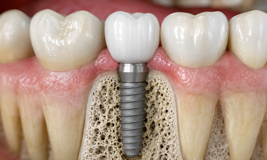
The Critical Role of Osseointegration
The placement of titanium dental implants represents the most critical surgical milestone in modern oral rehabilitation. These biocompatible fixtures essentially replace the missing natural root system. Following surgical insertion, the patient enters a mandatory healing phase, typically lasting three to six months. During this interval, the titanium posts undergo osseointegration.
This complex physiological process ensures the implants are permanently locked into the skeletal structure, capable of withstanding the immense biomechanical forces generated during active mastication (chewing).
The Sequence of Biological Stabilization
The clinical team must address three distinct anatomical challenges before any restorative prosthetics are fabricated. The following table outlines the pathological baseline conditions, the precise surgical interventions deployed, and the targeted clinical outcomes required to proceed.
| Pathological Baseline Condition | Targeted Clinical Intervention | Rehabilitated Clinical State (Outcome) |
| Active Periodontitis:
Deep gingival pockets harbor pathogenic bacterial colonies, leading to progressive alveolar bone resorption. |
Advanced Periodontal Therapy:
Execution of deep subgingival scaling, root planing, and laser-assisted curettage to remove calculus and necrotic tissue. |
Gingival Reattachment:
The complete cessation of localized inflammation results in firm, pink gingival tissue that tightly adheres to the root surfaces or implant abutments. |
| Non-Restorable Dentition:
Teeth exhibiting profound structural decay, severe Class III mobility, or persistent periapical lesions (abscesses). |
Strategic Surgical Extraction:
Meticulous, atraumatic removal of compromised teeth is frequently combined with immediate alveolar ridge preservation (bone grafting) within the socket. |
Pathology Eradication:
Complete removal of the infectious nidus and preservation of the necessary bone volume required to support future titanium anchors. |
| Generalized Edentulism (Missing Teeth):
Complete loss of the natural root structures, leading to a collapse of the occlusal vertical dimension. |
Implantology (Fixture Placement);
The surgical insertion of biocompatible endosseous titanium implants is precisely into the mapped alveolar bone. |
Osseointegration:
The physiological fusion of living osteoblasts directly to the titanium surface provides rigid, permanent biomechanical anchorage for the final superstructure. |
Only when the oral surgeon definitively confirms successful osseointegration and absolute periodontal stability can the patient advance to the final prosthodontic reconstruction phase. Building upon this meticulously prepared foundation guarantees the long-term predictability and functional longevity of the complete dental restoration.
The Restorative Phase and Rebuilding the Dentition
Following the successful osseointegration of the titanium fixtures and the complete healing of the periodontal tissues, the rehabilitation enters the restorative phase. This is the culmination of the treatment plan, where the prosthodontist fabricates and delivers the physical dental structures.
Unlike standard single-tooth dentistry, reconstructing an entire arch requires strict adherence to biomechanical engineering principles to ensure the new bite forces are distributed evenly across the jawbone. To break from conventional single-appointment restorative workflows, full mouth rehabilitation utilizes a strictly phased, data-driven progression to guarantee absolute phonetic, aesthetic, and functional success.
Phase I: Clinical Provisionalization (The Transitional Phase)
Before the final ceramic teeth are milled, the patient is fitted with a set of interim, or provisional, prosthetics.
These high-grade acrylic or composite restorations are temporarily fixed to the implant abutments or prepared natural teeth. This stage serves as a critical diagnostic test drive for the new oral architecture.
- Soft Tissue Conditioning: The contours of the provisional crowns actively shape the healing gingival tissue, creating a natural “emergence profile”, so the final teeth appear to grow organically from the gums.
- Occlusal Verification: The prosthodontist evaluates the patient’s new Vertical Dimension of Occlusion (VDO). This period allows the patient’s temporomandibular joint (TMJ) and masticatory muscles to adapt to the corrected jaw position.
- Phonetic Calibration: The structural shape of the anterior teeth directly dictates speech articulation. The provisional phase allows the clinical team to make immediate physical modifications to the acrylic if the patient experiences any phonetic interference (such as lisping or whistling).
The Importance of the Trial Period
The provisional phase typically lasts between four and eight weeks. The final ceramic prosthetics are never fabricated until the patient reports zero muscular fatigue, perfect speech articulation, and total masticatory comfort with the temporary restorations.
Phase II: Material Science and CAD/CAM Fabrication
Once the provisional architecture is clinically validated, the exact spatial data is transferred to the dental laboratory via digital intraoral scanners. The fabrication of the final prosthesis leverages advanced Computer-Aided Design and Computer-Aided Manufacturing (CAD/CAM) technology. The selection of restorative materials is dictated strictly by the required tensile strength and aesthetic demands of the specific anatomical region.
- Monolithic Zirconia:
Utilized primarily for full-arch, implant-supported bridges and posterior crowns.
Zirconia is a highly biocompatible ceramic boasting immense flexural strength, making it completely resistant to the heavy biomechanical forces generated during mastication, while preventing catastrophic fractures.
- Lithium Disilicate (E.max):
Frequently deployed for anterior (front) restorations, such as custom crowns and veneers.
This glass-ceramic provides superior optical translucency, mimicking the exact light-reflecting properties of natural human enamel.
- Titanium Frameworks:
For patients requiring complete upper or lower arch replacements (hybrid prostheses), a computer-milled titanium bar is often embedded within the ceramic or acrylic structure.
This provides rigid, unyielding skeletal support across the entire span of the jaw.
Phase III: Delivery and Occlusal Equilibration
The final clinical appointment involves the permanent fixation of the restorative superstructures. However, simply attaching the prosthetics is insufficient. The prosthodontist must perform an occlusal equilibration.
As natural teeth possess a periodontal ligament that provides slight micromovement and proprioception (pressure sensing), whereas titanium implants are rigidly locked into the bone, the bite must be calibrated with microscopic precision.
The specialist utilizes articulating foils and digital T-scan sensors to identify and eliminate any premature contact points.
This ensures that the masticatory forces are dispersed symmetrically across the entire arch, preventing targeted mechanical overload and guaranteeing the multi-decade longevity of the full mouth rehabilitation.
The Clinical Benefits of Rehabilitation
While the aesthetic transformation following a complete oral reconstruction is profound, the primary medical objectives of this intervention are strictly physiological. Chronic edentulism and severe occlusal degradation trigger a cascade of systemic health complications that extend far beyond the oral cavity.
Definitively restoring the structural integrity of the maxillofacial region, full mouth rehabilitation yields critical, long-term systemic benefits. Clinical specialists categorize these physiological outcomes into three primary domains of patient health:
Gastrointestinal and Nutritional Optimization
The oral cavity serves as the primary gateway for the human digestive system. When the mechanical grinding capability of the teeth is compromised, the entire gastrointestinal tract suffers functional overload.
- Masticatory Force Restoration: Traditional removable dentures only provide a fraction of natural chewing power. Biomechanically anchored titanium implants restore masticatory force to near natural baseline levels, allowing patients to confidently consume fibrous, nutrient-dense foods.
- Enzymatic Processing: Proper mechanical breakdown (comminution) of food within the oral cavity is an absolute biological requirement for the activation of salivary amylase.
- Nutrient Absorption: By ensuring food is adequately pulverized before swallowing, the rehabilitated dentition directly enhances gastrointestinal absorption, actively preventing the systemic malnutrition and digestive distress frequently observed in patients with compromised bites.
Craniofacial and Neuromuscular Stabilization
A collapsed bite fundamentally alters the anatomical resting position of the lower jaw, placing immense, destructive strain on the surrounding skeletal and muscular architecture.
| Anatomical Region | Pathological Degraded State | Rehabilitated Clinical State |
| Temporomandibular Joint (TMJ) | Chronic subluxation, severe muscular fatigue, and persistent tension headaches caused by a collapsed vertical dimension of occlusion. | The newly engineered prosthetics re-establish the correct vertical height, physically decompressing the joint space and resolving chronic facial pain. |
| Alveolar Bone (Jawbone) | Progressive anatomical atrophy and volumetric resorption triggered by the absence of natural tooth roots. | Endosseous titanium implants provide constant internal biomechanical stimulation to the osteoblasts, permanently halting bone loss. |
| Maxillofacial Musculature | Muscular hypertrophy or pathological spasms resulting from the jaw constantly overcompensating for an imbalanced, asymmetrical bite. | Symmetrical occlusal contacts distribute bite forces evenly, allowing the complex masticatory muscles to return to a relaxed, natural baseline tone. |
Phonetic and Structural Preservation
The teeth provide critical structural support for the entire lower third of the human face. Their absence or severe degradation leads to immediate phonetic impairment and premature facial aging.
- Articulatory Precision: The precise spatial positioning of the new anterior ceramic restorations provides the exact structural boundaries required by the tongue and lips to form complex phonetic sounds, permanently correcting speech impediments such as lisping or slurring.
- Facial Profile Support: Rebuilding the proper vertical height of the teeth physically supports the perioral musculature. This structural reinforcement instantly reverses the sunken, collapsed facial appearance heavily associated with advanced tooth loss.
- Psychological Restoration: Completing a definitive reconstruction eliminates the chronic psychological burden of failing dentition, the fear of prosthetic dislodgement, and the pain of active oral infections.
Addressing the entire masticatory system comprehensively, then full mouth rehabilitation transitions the patient from a state of chronic physiological decline to a state of absolute, stabilized oral function.
Why Choose DDC Smiles for Complex Oral Reconstruction?
Executing a complete oral reconstruction requires an institutional framework capable of supporting advanced surgical and prosthodontic interventions. DDC Smiles operates as a premier dental hospital, providing the specific multidisciplinary environment required for highly predictable full-arch restorations in Bangalore.
The institution guarantees clinical excellence through several highly specific structural and operational advantages:
- In-House 3D Diagnostics: The facility utilizes on-site Dental CBCT (PaX-i3D) and Digital OPG technology to acquire exact volumetric data. This capability allows the surgical team to map critical neurovascular pathways immediately, eliminating the need for external anatomical mapping.
- Multidisciplinary MDS Board: Full mouth rehabilitation requires diverse clinical expertise. The institution houses a collaborative team of over twelve credentialed specialists, directed by Dr. H.J. Jaikrishna, who ensures every surgical and restorative phase is executed by a dedicated domain expert.
- Hospital-Grade Sterilization Protocols: Extensive implant surgery requires absolute biological safety. The clinical staff strictly adheres to a six-step sterilization pathway utilizing Class B Autoclaves, maintaining a pristine, infection-free surgical environment.
With over twenty-three years of dedicated institutional experience and thousands of successful implant placements, the facility possesses the specific empirical knowledge required to manage and resolve complex structural failures.
Conclusion
Severe structural degradation of the oral cavity is a completely reversible medical condition. By systematically combining advanced periodontics, precision implantology, and custom prosthodontics, full mouth rehabilitation fully restores both physiological function and anatomical integrity.
Patients experiencing advanced dental collapse or generalized tooth loss should not accept permanent functional impairment. Schedule a comprehensive diagnostic evaluation with the clinical specialists at DDC Smiles to precisely map your anatomy and initiate your specific structural reconstruction.
