


Experiencing persistent dental discomfort often makes people suspect a deep cavity, but underlying teeth pain gum disease connections are among the most overlooked causes of chronic dental aches.
When seeking care at a specialized Dental Hospital in HSR Layout and Koramangala, accurate diagnostic evaluation reveals that inflamed supporting periodontal tissues directly irritate the neuro-vascular pathways surrounding the tooth root.
Teeth do not sit isolated in the jaw; they are anchored by the periodontal ligament (PDL) a shock-absorbing connective tissue matrix rich in nerve endings and pressure-sensing receptors.
When bacterial plaque extends below the gumline into the gingival sulcus, inflammatory swelling exerts localized pressure on the tooth root, perfectly mimicking a traditional toothache.
What You Will Discover in This Guide
- Anatomical Comparison: Structural differences between healthy, inflamed, and compromised gum tissue.
- The 7 Hallmark Warning Signs: Deconstructing specific pain patterns, from chewing tenderness to root exposure.
- The Endo-Perio Dilemma: How to tell if pain originates from a dying tooth nerve or a deep periodontal pocket.
- Targeted Clinical Solutions: Advanced 3D imaging and minimally invasive treatments to eliminate pain at its biological source.
Addressing dental pain requires treating the entire supporting foundation. At DDC Smiles, our multi-specialty team combines low-dose 3D digital imaging with targeted periodontal therapy to diagnose and resolve the root cause of your discomfort.
Clinical Dental Disclaimer
The diagnostic comparisons, anatomical pathways, and clinical symptom descriptions presented in this guide are intended strictly for educational and public health awareness purposes. This content does not constitute formal dental advice, clinical diagnosis, or a personalized restorative treatment plan. Accurately diagnosing the underlying cause of tooth or gum pain requires an in-person clinical examination, vitality testing, and radiographic imaging by a licensed dentist, endodontist, or periodontist.
Anatomical Comparison: Tissue Architecture & Warning Markers
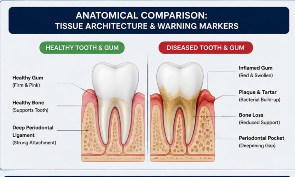
Understanding why periodontal issues manifest as tooth pain requires examining the structural integrity of the surrounding periodontium.
When comparing healthy gums vs gingivitis and advanced periodontitis, the transition from tight, protective tissue to chronically inflamed, permeable pockets alters the mechanical support of the tooth, exposing sensitive root architecture and triggering nerve irritation.
3-Tier Periodontal Architecture Matrix
| Clinical Parameter | Stage 1: Healthy Periodontium | Stage 2: Gingivitis (Reversible) | Stage 3: Periodontitis (Irreversible Bone Loss)
|
|---|---|---|---|
| Gingival Margin & Color | Firm, stippled, pale coral-pink; knife-edged margins tightly hugging the tooth enamel. | Erythematous (bright red), edematous (swollen), smooth, and glossy with loss of stippling. | Dusky red, purplish, or cyanotic; receded gingival margins exposing the underlying root dentin. |
| Sulcular Depth (Pocket Probing) | 1 to 3 mm (shallow, cleansable, with an intact junctional epithelium). | 2 to 4 mm (“pseudo-pockets” caused solely by soft tissue swelling without bone loss). | 4 to 9+ mm (true pathological pockets with apical migration of junctional epithelium). |
| Bleeding On Probing (BOP) | Negative; zero bleeding during routine brushing, flossing, or diagnostic periodontal probing. | Positive; capillaries are dilated and micro-ulcerated, bleeding easily on gentle contact. | Chronic, spontaneous bleeding accompanied by purulent exudate (pus) upon gentle pressure. |
| Periodontal Ligament (PDL) & Bone | Intact alveolar crest; dense Sharpey’s fibers firmly anchoring root cementum to alveolar bone. | Intact alveolar bone levels; inflammation is strictly confined to the superficial epithelial layers. | Progressive destruction of PDL fibers and horizontal/vertical resorption of supporting alveolar bone. |
| Tooth Stability & Pain Profile | Zero mobility; teeth comfortably absorb normal masticatory (chewing) forces without ache. | Minimal direct discomfort; occasional localized tenderness or itching along the gumline. | Pathological tooth mobility, dull throbbing ache, bite sensitivity, and sharp pain on temperature changes. |
How Structural Breakdown Triggers Dental Pain
- Loss of the Epithelial Seal: In a healthy state, the junctional epithelium acts as a biological barrier. As bacterial toxins degrade this seal, bacteria travel down the root surface, stimulating nociceptors (pain receptors) inside the periodontal ligament.
- Microvascular Engorgement: Inflammatory mediators (prostaglandins and interleukins) cause local blood vessels to dilate and leak fluid into the enclosed space around the tooth root, building up localized hydrostatic pressure.
- Dentin Exposure via Recession: When chronic inflammation causes the gum margin to recede, the thin, protective cementum layer on the root wears away. This exposes porous dentinal tubules, which conduct hot, cold, and acidic stimuli directly to the internal dental pulp.
The Diagnostic Takeaway: Gingivitis is entirely reversible As the supporting bone and periodontal ligament remain intact. Once inflammation crosses the threshold into periodontitis, structural bone loss begins, converting simple gum sensitivity into deep-seated toothache.
The 7 Hallmark Signs
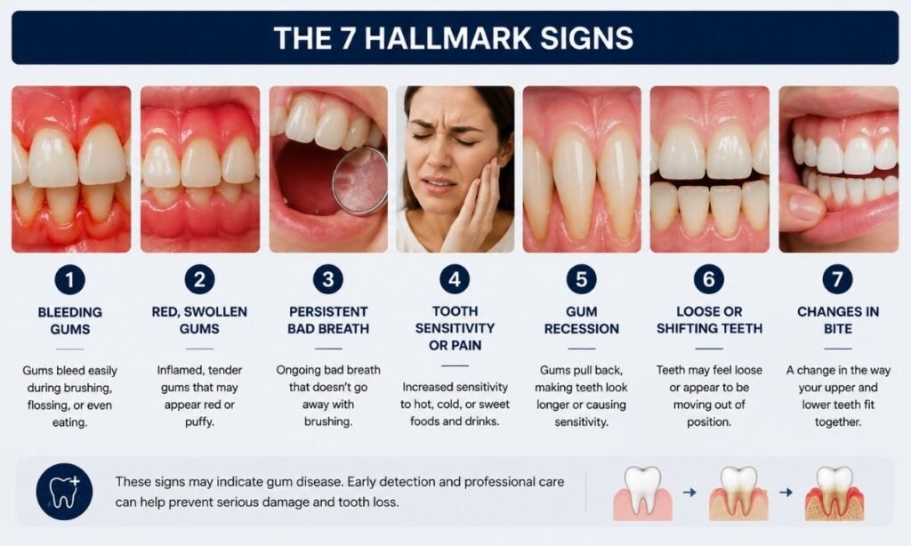
Dental discomfort resulting from periodontal breakdown has a distinct clinical presentation compared to a localized enamel cavity. When evaluating gum disease and symptoms that manifest as dental pain, the issue rarely stems from sudden enamel decay alone.
Instead, pain reflects progressive inflammation of the periodontal ligament, root cementum exposure, or localized subgingival pressure.
The 7 Warning Signs Your Tooth Pain Stems from Gum Disease
- A Persistent, Dull, Deep-Seated Aching in the Jawbone
The Mechanism: Unlike the sharp, electric twinge of a superficial cavity, inflamed periodontal fibers produce a continuous, low-grade ache deep within the alveolar bone. As the periodontal ligament is dense with mechanoreceptors, inflammation registers as a diffused, throbbing soreness across an entire quadrant.
Clinical Context: Often described as a “tired” or “heavy” sensation around the back molars that worsens toward the end of the day.
- Sharp Discomfort When Biting Down or Chewing
The Mechanism: Chewing forces push the tooth slightly into its socket. When the surrounding periodontal ligament is swollen and engorged with inflammatory fluid, this normal downward movement compresses the irritated nerve fibers directly against the bony socket wall.
Clinical Context: Patients often instinctively chew on the opposite side of their mouth to avoid the mechanical pressure on the affected tooth.
- Sudden Sensitivity to Hot, Cold, or Sweet Stimuli
The Mechanism: As chronic inflammation forces the gingival margin to recede, the protective enamel boundary is lost, exposing the tooth’s root. The microscopic dentinal tubules on the root surface lead straight to the inner pulp, allowing thermal and osmotic changes to trigger immediate sensitivity.
Clinical Context: Pain flares up sharply when drinking iced water, hot tea, or eating acidic foods, even if dental radiographs show zero cavities in the crown.
- Localized Tension or Pressure Between Two Specific Teeth
The Mechanism: Deep periodontal pockets create sheltered voids where fibrous food particles and bacterial biofilm become trapped. The resulting localized immune response triggers acute swelling inside the confined interdental space.
Clinical Context: Temporary relief is often felt immediately after flossing or picking between the teeth, but the deep pressure and tenderness quickly return.
- The Sensation of a “High,” Loose, or Floating Tooth
The Mechanism: As supporting alveolar bone resorbs and inflammatory edema expands inside the socket, the tooth is micro-elevated out of its natural resting position. When closing the mouth, this tooth hits the opposing arch prematurely.
Clinical Context: The tooth feels slightly mobile, unstable, or out of alignment when chewing, leading to secondary occlusal trauma.
- A Constant Metallic Taste or Foul Odor Accompanying the Ache
The Mechanism: Anaerobic bacteria flourishing in deep, oxygen-depleted pockets produce volatile sulfur compounds (VSCs) and purulent exudate (microscopic pus) that seeps into the oral cavity.
Clinical Context: Persistent halitosis and an unpleasant salty or bitter taste accompany the toothache, unaffected by standard mouthwashes or mints.
- Throbbing Pain That Intensifies When Lying Flat
The Mechanism: When shifting from an upright position to lying down at night, systemic blood pressure redistributes, increasing cephalic and microvascular blood flow to the head and oral cavity. This creates heightened hydrostatic pressure inside already engorged periodontal pockets.
Clinical Context: Tooth throbbing awakens patients from sleep or makes falling asleep difficult, frequently mistaken for an acute root canal emergency.
The Diagnostic Clue: If a hurting tooth feels sore to tap, sensitive to cold at the gumline, and is surrounded by gums that bleed when brushed, the underlying cause is almost certainly periodontal rather than a simple enamel cavity.
The Pathological Progression Curve: From Reversible to Irreversible
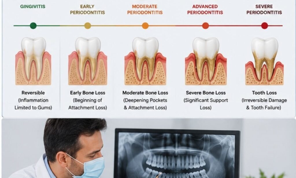
The journey from mild plaque accumulation to debilitating periodontal toothache is not an overnight event; it follows a predictable biological cascade driven by bacterial dysbiosis and host immune responses.
Catching and treating early gum disease during its initial stages is vital As superficial gingival inflammation is completely reversible, whereas unmanaged infection triggers irreversible alveolar bone loss and permanent tissue attachment damage.
Biological Staging Progression Flow
| The Periodontal Disease Progression
|
|---|
| Phase 1: Biofilm Infiltration (0–4 Days) ➔ Phase 2: Early Gingivitis & Sulcular Ulceration (4–14 Days) [CRITICAL WINDOW OF REVERSIBILITY] ➔ Phase 3: Established Pocketing & Periodontitis Onset (Sub-acute) ➔ Phase 4: Advanced Periodontitis & Osteoclast Bone Resorption (Chronic) |
Phase 1: Microbial Biofilm Colonization (Days 0 to 4)
- Biological Mechanism: Salivary glycoproteins form an acquired pellicle on clean enamel. Aerobic gram-positive bacteria (Streptococcus sanguinis, Actinomyces) adhere, rapidly proliferating into an organized sticky matrix (dental plaque).
- Tissue Impact: Gingival tissues appear clinically normal, but microscopic chemotaxis begins as neutrophils migrate toward the gingival sulcus to neutralize early bacterial antigens.
Phase 2: Early Gingival Breakdown & Vascular Dilation (Days 4 to 14)
- Biological Mechanism: As plaque thickens and oxygen levels drop, colonies transition to facultative and gram-negative anaerobes. Endotoxins irritate the sulcular lining, triggering localized histamine and prostaglandin release.
- Clinical Presentation: Gums become red, puffy, and bleed upon contact. Micro-ulcerations form on the inner pocket wall, creating microscopic entry points for bacteria.
- Reversibility Window: 100% Reversible. Professional dental scaling combined with diligent daily flossing fully restores healthy collagen architecture without permanent structural loss.
Phase 3: True Pocket Formation & Periodontal Ligament Detachment (Sub-Acute)
- Biological Mechanism: Chronic immune activation leads to matrix metalloproteinases (MMPs) breaking down the Sharpey’s fibers of the periodontal ligament. The junctional epithelium detaches from the enamel and migrates downward along the root.
- Clinical Presentation: Sulcular depths exceed 4 mm, forming sheltered, anaerobic “true pockets.” Patients begin experiencing localized tenderness, food impaction, and bite sensitivity as fluid pressure builds inside the confined pocket.
Phase 4: Osteoclast Activation & Irreversible Bone Resorption (Chronic Periodontitis)
- Biological Mechanism: Pro-inflammatory cytokines (RANKL, TNF-α, IL-1β) stimulate osteoclasts cells that resorb and destroy bone tissue. Alveolar bone recedes away from the infection front to protect deeper skeletal structures.
- Clinical Presentation: Tooth mobility, persistent deep throbbing aches, visible gum recession with exposed root dentin, and potential periodontal abscess formation. Once alveolar bone height is lost, it cannot regenerate spontaneously without advanced surgical grafting or regenerative laser therapies.
The Clinical Reality: Tooth pain rarely appears during the painless, reversible phases of gingivitis. By the time a patient feels deep chewing soreness or throbbing pressure, periodontal disease has already advanced into the bone-destructive stages.
The Endo-Perio Dilemma (Pulp vs. Gum Origin)
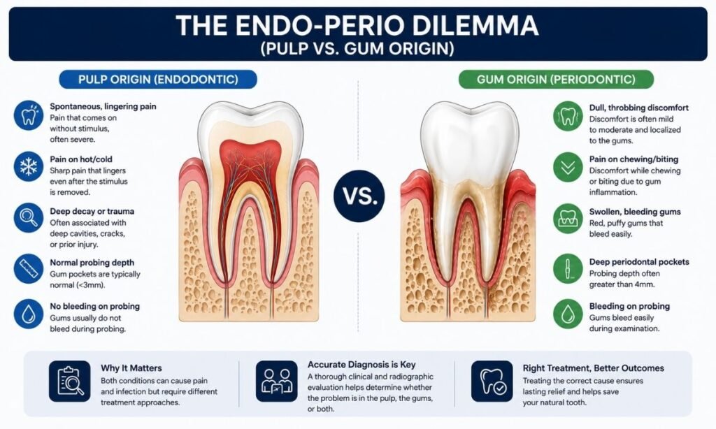
Determining the true origin of dental pain is one of the most critical diagnostic challenges in clinical dentistry.
As the internal dental pulp and the surrounding periodontium share anatomical pathways including the apical foramen, lateral accessory canals, and exposed dentinal tubules infections can travel bidirectionally between the tooth’s nerve and its supporting bone.
Differentiating pulpal necrosis from advanced gum disorders ensures that patients receive the correct targeted treatment, avoiding unnecessary root canals when the true issue is periodontal, or vice versa.
Diagnostic Decision Matrix: Pulpal vs. Periodontal Pain Origin
| Clinical Parameter | Primary Endodontic Lesion (Nerve / Pulp Origin) | Primary Periodontal Lesion (Gum / Bone Origin) | True Combined Endo-Perio Lesion (Dual Origin)
|
|---|---|---|---|
| Pain Character & Localization | Sharp, shooting, or intense throbbing; often poorly localized and can radiate across the jaw. | Dull, continuous ache; accurately localized to a specific tooth with a feeling of deep pressure. | Complex, severe throbbing combined with continuous pressure and bite tenderness. |
| Thermal Sensitivity (Cold / Heat) | Exaggerated, lingering response to cold/heat, or complete absence of sensation (non-vital pulp). | Normal response to thermal tests; root sensitivity is brief and subsides immediately upon removal of stimulus. | Non-responsive to vitality tests; cold/heat does not register due to pulpal necrosis. |
| Electric Pulp Testing (EPT) | Negative (no response), indicating non-vital or necrotic pulpal tissue. | Positive (normal vital response), confirming that the internal nerve remains healthy. | Negative (no response); both internal nerve tissue and surrounding periodontium are infected. |
| Percussion (Tapping Test) | Severe, sharp pain when tapping the biting surface vertically. | Moderate tenderness when tapping the tooth horizontally against the gumline. | Severe pain to both vertical and horizontal percussion. |
| Periodontal Probing Depth | Typically isolated, narrow, single-point deep probing defect at the root apex. | Wide, generalized deep pocketing (>5 mm) spanning multiple tooth surfaces with calculus. | Deep, wide, generalized pocket extending all the way to the root apex. |
| Radiographic Appearance | Localized radiolucency (dark shadow) confined to the root tip (periapical area). | Horizontal or angular crestal bone loss originating at the gumline and moving downward. | Continuous bone destruction connecting the crestal gum margin directly to the root apex. |
The Pathways of Cross-Infection
When periodontal or endodontic conditions go unmanaged, bacteria exploit three primary anatomical bridges to spread infection:
- 1. Apical Foramen: Primary exit for deep pulpal necrosis into bone.
- 2. Lateral & Accessory Canals: Micro-conduits along the root mid-body.
- 3. Dentinal Tubules: Porous micro-channels exposed by gum recession.
- Retrograde Pulpitis (Gums ➔ Nerve): Severe periodontal pockets extending down the root allow subgingival anaerobic pathogens to enter lateral canals or the apical foramen, secondarily infecting an otherwise healthy, cavity-free dental pulp.
- Secondary Periodontal Breakdown (Nerve ➔ Gums): A necrotic, infected pulp leaks toxins through the apical foramen into the surrounding periodontal ligament, mimicking a deep gum pocket and causing rapid alveolar bone breakdown from the inside out.
A tooth should never undergo an irreversible root canal based on pain alone. Comprehensive diagnostic testing including electric pulp vitality assessments, 3D bone mapping, and multi-point periodontal probing is essential to isolate whether the infection stems from the internal nerve or the external periodontal foundation.
Why Choose DDC Smiles? 3D Mapping & Targeted Care
Resolving complex dental pain requires moving beyond guesswork to isolate the exact biological source of discomfort. When consulting an experienced Dentist in HSR Layout, patients at DDC Smiles benefit from an advanced diagnostic infrastructure designed to differentiate between pulpal nerve damage and periodontal bone loss, ensuring treatments target the actual root cause rather than just masking symptoms.
Key Clinical Pillars at DDC Smiles
- Low-Dose 3D CBCT Bone & Root Imaging: Standard two-dimensional X-rays often superimpose overlapping anatomical structures, masking early bone resorption or lateral canal infections. Our high-resolution Cone Beam Computed Tomography (CBCT) provides clear 3D volumetric mapping of alveolar bone height, root morphology, and periapical tissues with sub-millimeter precision.
- Multi-Specialty Endo-Perio Diagnostic Synergy: As pain pathways between the tooth nerve and the surrounding periodontium frequently overlap, every complex case is co-evaluated by our endodontic and periodontal specialists. Combining electric pulp vitality testing, thermal mapping, and digital periodontal probing prevents unnecessary root canals and ensures appropriate gum therapy.
- Dual-Wavelength Photonic Laser Therapy: For patients experiencing deep pocket inflammation and bone loss, our clinic utilizes advanced soft-tissue Diode and hard-tissue Erbium lasers. Photonic decontamination eliminates subgingival microbial biofilms, degranulates diseased tissue, and biostimulates periodontal ligament healing without scalpel incisions or surgical sutures.
- Minimally Invasive Microscopic Restorations: When gum recession exposes sensitive root dentin, our restorative specialists apply biocompatible composite resins and glass ionomers under high-magnification dental microscopes, instantly sealing open dentinal tubules and restoring smooth, cleansable contours along the gumline.
- Stress-Free, Patient-Centric Care: We understand that toothache often carries significant physical discomfort and anxiety. Our gentle, vibration-free laser protocols, needle-free numbing options, and supportive environment ensure your visit is comfortable and pain-free from start to finish.
The DDC Smiles Pain-Resolution Protocol
- Digital Diagnostic Mapping: High-definition intraoral cameras and 3D CBCT scans identify the exact structural or periodontal defect.
- Targeted Biological Intervention: Whether requiring laser pocket sterilization, microscopic root canal therapy, or cervical bonding, care is tailored to preserve your natural tooth.
- Preventive Stabilization Plan: Structured follow-ups and periodontal maintenance protect the surrounding bone and prevent recurring discomfort.
The DDC Smiles Standard: Tooth pain is a symptom, not a final diagnosis. By combining advanced 3D imaging with multi-specialty expertise, we identify the true origin of your discomfort and deliver targeted, tooth-preserving solutions.
Conclusion
Masking dental pain with temporary painkillers only allows subgingival bacterial infections and bone loss to progress silently beneath the gumline. As tooth pain is frequently a secondary symptom of underlying periodontal breakdown, getting a comprehensive clinical evaluation from a trusted Dentist in Koramangala or neighboring HSR Layout ensures the true origin of your discomfort is identified and treated before permanent bone loss occurs.
Through advanced 3D digital imaging, electric pulp vitality assessments, and precise periodontal mapping, our dental specialists isolate whether your pain stems from an internal root canal infection or an external gum pocket delivering targeted, conservative care designed to save your natural teeth.
Schedule Your Diagnostic Evaluation at DDC Smiles
If you are experiencing persistent tooth tenderness, chewing sensitivity, swollen gums, or unexplained dental throbbing, contact our multi-specialty team in South Bengaluru to book an appointment:
- Clinic Address: DDC Smiles Dental Clinic & Implant Centre, HSR Layout / Koramangala Sector, Bengaluru, Karnataka
- Appointment Booking & Inquiries: Connect directly via ddcsmiles.in
- Website: ddcsmiles.in
Specialized Dental Services Available: Periodontal Pocket Therapy & Gum Care, Microscopic & Rotary Endodontics (Painless RCT), Dual-Wavelength Laser Dentistry, Dental Implants (Single, Multiple & Full-Arch), Clear Aligners, and Cosmetic Smile Design.
Reference:
https://www.columbiadoctors.org/health-library/symptom/toothache-gum-problems/
