
Whether arriving at a dental clinic, the speed of the medical response strictly dictates tooth survival. As a premier Dental Hospital in Koramangala and an advanced Dental Hospital in HSR Layout, DDC Smiles utilizes on-site Digital OPG technology to instantly assess severe structural damage.
During these critical moments, patients naturally ask exactly what dental trauma is and how specialized emergency dentistry can save compromised teeth.
Unlike routine dental decay, which progresses slowly over months, a traumatic injury causes instantaneous, catastrophic disruption to the hard tissues, the internal dental pulp, and the surrounding supportive bone.
In these severe scenarios, time is the absolute most critical biological factor. The probability of successfully reimplanting an avulsed tooth or stabilizing a severe fracture drops precipitously with every passing minute.
To provide complete clinical clarity for patients and parents navigating these highly stressful emergencies, this guide will detail the strict medical protocols governing dental traumatology. We will systematically examine:
1. The Clinical Scope: Defining the specific physical forces involved in these injuries and the need for immediate specialised intervention.
2. Injury Classification: Detailing how clinicians categorically assess the severity of the damage, ranging from minor enamel chipping to complete anatomical displacement.
3. Restorative Treatment Protocols: Explaining the precise surgical and endodontic procedures used to stabilize the injured tooth and preserve long-term oral function.
Understanding the critical urgency and specific medical protocols associated with maxillofacial injuries enables patients to respond to emergencies with rapid, decisive action to ensure the best possible clinical outcome.
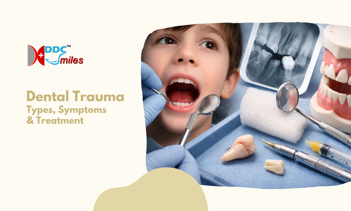
What is Dental Trauma?
When a patient questions what dental trauma is, the clinical definition extends far beyond a simple chipped tooth.
In maxillofacial medicine, this term strictly encompasses any acute physical injury sustained by the hard dental tissues, the internal neurovascular pulp, the supporting periodontal structures, or the adjacent oral soft tissues.
These injuries occur when a sudden, high-velocity physical force impacts the facial skeleton, transferring severe kinetic energy directly into the complex biological architecture of the mouth.
To accurately assess the total scope of a traumatic impact, clinical specialists systematically evaluate four distinct anatomical zones:
1. Hard Dental Tissues: This includes the external enamel crown, the underlying dentin layer, and the cementum covering the root surface. High-impact forces frequently cause structural fractures within these highly dense crystalline layers.
2. The Dental Pulp: The innermost chamber of the tooth contains a sensitive matrix of nerves and blood vessels. Blunt force trauma can completely sever the blood supply at the root apex, causing immediate pulpal necrosis even if the external tooth remains structurally intact.
3. The Periodontal Apparatus: This crucial anatomical support system consists of the periodontal ligament and the surrounding alveolar bone socket. Severe impacts can crush the bony socket or violently stretch the ligament, resulting in tooth displacement or complete avulsion.
4. Oral Soft Tissues: The lips, internal buccal mucosa, and gingival tissues frequently sustain deep lacerations or severe contusions during the primary impact, requiring immediate surgical suturing.
As these emergencies involve complex, deeply interconnected biological structures, their precise medical management falls under the dedicated clinical discipline of Dental Traumatology.
Specialists within this field do not merely focus on cosmetic tooth restoration. Their primary clinical objective is to rapidly stabilize the injured structures, reestablish the disrupted neurovascular blood supply, and prevent the onset of aggressive biological root resorption or severe bone infections following the initial traumatic event.
Dental Trauma Classification
Clinicians do not treat all facial injuries uniformly. The specific surgical or endodontic approach depends entirely on a highly standardized assessment of the physical damage.
This precise clinical framework, known universally as the dental trauma classification system, allows specialists to rapidly evaluate the exact nature of the injury and immediately initiate the correct emergency protocol.
Clinicians categorize these injuries based on the specific anatomical tissues compromised during the impact. The severity ranges from superficial structural loss to catastrophic displacement.
Standard Clinical Classifications of Dental Injuries
| Injury Classification | Clinical Definition | Primary Anatomical Involvement |
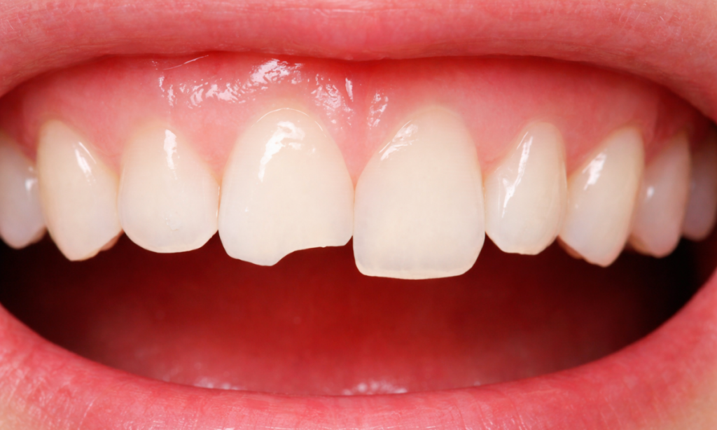
| Uncomplicated Crown Fractures | A structural break is isolated strictly to the outer layers of the tooth. | The highly mineralized enamel and the underlying dentin, with absolutely no exposure of the internal pulp chamber. |
| Complicated Crown Fractures | A severe structural break that physically breaches the innermost chamber of the tooth. | The enamel, the dentin, and the direct exposure of the highly sensitive neurovascular dental pulp to the external oral environment. |
| Root Fractures | A horizontal or diagonal break occurring entirely below the gum line. | The cementum, dentin, and pulp are located specifically within the alveolar bone socket. These strictly require high-resolution radiographic imaging for detection. |
| Luxation (Displacement) | The tooth remains physically within the mouth but is violently forced out of its normal anatomical alignment. | Severe tearing or stretching of the periodontal ligament. The tooth may be pushed deeper into the bone (intrusion), pulled partially out (extrusion), or pushed sideways (lateral luxation). |
| Complete Avulsion | The tooth is entirely knocked out of the alveolar bone socket. | Total severance of the periodontal ligament, the apical blood supply, and the primary nerve bundle. This is the most severe clinical emergency in dental traumatology. |
Accurate classification is the absolute foundation of emergency dental trauma management. By immediately identifying the exact type of structural and periodontal damage, the clinical team can prevent irreversible tissue necrosis, stop aggressive bone infections, and significantly increase the probability of successfully retaining the natural tooth structure.
Dental Trauma in Children
Dental injuries in pediatric patients demand a fundamentally different clinical approach compared to adult emergencies.
When evaluating dental trauma in children, the surgical team must account for a highly complex biological variable: the primary dentition physically resides directly above the developing permanent dentition, currently hidden deep within the alveolar bone.
Treating a pediatric dental injury is never strictly about repairing the visible baby tooth. The primary clinical objective is to protect the unerupted adult tooth beneath it. Pediatric oral structures possess several unique anatomical vulnerabilities that dictate emergency treatment protocols:
1. Extreme Anatomical Proximity: The root apex of a primary incisor sits mere millimeters away from the delicate, developing follicle of the permanent adult incisor. Any severe physical force directed at the primary tooth is instantly transferred to the permanent tooth bud.
2. Alveolar Bone Pliability: Pediatric jawbones are highly pliable and significantly less dense than adult bone. Consequently, high velocity impacts frequently cause severe tooth displacement, forcing the tooth deep into the bone, rather than causing a clean structural crown fracture.
3. Incomplete Root Formation: When a newly erupted permanent tooth sustains severe trauma, the root structure is often immature, and the apex remains physically open. Saving these teeth requires highly specialized endodontic procedures, such as apexification, to artificially stimulate the root to finish developing and seal completely.
As of this anatomical proximity, specific injuries to primary teeth carry a high statistical probability of causing permanent, irreversible developmental defects in the adult teeth.
Clinical Consequences of Primary Tooth Trauma
| Primary Tooth Injury | Physical Mechanism of Damage | Potential Consequences for the Unerupted Permanent Tooth |
| Severe Intrusion | The primary tooth is violently driven upward directly into the alveolar bone, physically crushing the underlying permanent tooth follicle. | Enamel hypoplasia (severe structural discoloration), severe crown malformation, or root dilaceration (abnormal bending of the adult root). |
| Complete Avulsion | The primary tooth is entirely knocked out. Note: Primary teeth are absolutely never reimplanted due to the extreme risk of damaging the adult tooth during the reimplantation process. | Delayed eruption of the adult tooth or severe ectopic (anatomically misaligned) eruption requiring future orthodontic surgical intervention. |
| Untreated Pulpal Infection | Following a minor, ignored trauma, the primary tooth nerve dies. Chronic bacterial infection spreads from the dead root apex directly into the surrounding bone. | Localized arrest of permanent tooth development or severe enamel defects is clinically known as Turner’s hypoplasia. |
Utilizing high-resolution pediatric radiography immediately following an injury, the clinical team can precisely map the trajectory of the trauma.
This ensures that the emergency intervention stabilizes the immediate damage while strictly preserving the biological development of the patient’s future permanent smile.
Dental Trauma Treatment
When a patient arrives at the emergency dental suite following a facial injury, the immediate clinical objectives are highly specific: physically stabilize the mobile dentition, seal the internal pulp chamber from bacterial contamination, and restore normal masticatory function.
The specific dental trauma treatment selected depends entirely on the exact classification of the structural damage and the biological maturity of the tooth root.
Specialized endodontists utilize a strict hierarchy of restorative and surgical protocols to manage these complex emergency scenarios, ensuring accurate stabilization and long-term preservation of injured teeth.
Clinical Intervention Matrix for Dental Injuries
| Specific Injury Classification | Primary Surgical or Restorative Protocol | Primary Biological Objective |
| Uncomplicated Crown Fracture | Direct composite resin bonding or autogenous fragment reattachment (bonding the original broken tooth piece back into place). | To hermetically seal the exposed dentinal tubules, preventing severe thermal sensitivity and restoring the original anatomical contour of the crown. |
| Complicated Crown Fracture | Vital pulp therapy (partial pulpotomy) or complete root canal therapy, followed immediately by a full coverage ceramic crown. | To surgically excise the superficially contaminated nerve tissue while aggressively attempting to preserve the healthy, sterile pulp situated deeper within the root canal system. |
| Luxation (Severe Displacement) | Manual or forceps repositioning followed immediately by the application of a flexible wire and composite splint for two to four weeks. | To physically immobilize the traumatized tooth within the arch, allowing the torn periodontal ligament fibres adequate time to biologically reattach to the alveolar bone socket. |
| Complete Avulsion (Knocked Out) | Immediate surgical reimplantation back into the anatomical socket, flexible splinting, and mandatory subsequent endodontic therapy. | To reestablish the tooth within the bone. The subsequent root canal is absolutely mandatory because the apical blood supply is permanently severed upon complete avulsion. |
Advanced Endodontic Considerations
The most complex aspect of managing severe dental impacts involves the internal neurovascular pulp. When an injury physically severs the blood supply at the root apex, the internal tissue inevitably undergoes necrosis (cellular death).
To prevent the rapid onset of severe periapical abscesses and aggressive bone infections, the clinical team must execute precise endodontic interventions based specifically on the developmental stage of the root structure:
1. Mature Roots (Closed Apex): For adult patients with fully developed teeth, the endodontist performs standard root canal therapy.
They chemically disinfect the internal chamber and mechanically seal the entire canal system with a biocompatible filling material to permanently prevent deep bacterial colonization.
2. Immature Roots (Open Apex): In pediatric and adolescent patients, standard root canal therapy is strictly contraindicated because the root walls are physically thin and structurally weak.
The specialist instead utilizes advanced procedures such as apexification. This complex intervention involves placing a specialized bioactive cement directly at the open root tip. This precise material artificially induces the formation of a hard tissue calcific barrier, successfully sealing the root and preserving the fragile tooth structure.
Strictly adhering to these advanced surgical and endodontic protocols, the clinical team maximizes the statistical probability of long-term tooth survival and prevents the severe biological complications associated with untreated facial injuries.
Why Choose DDC Smiles?
When a severe dental injury occurs, navigating between a general dental clinic and a separate external diagnostic center wastes critical biological time.
At DDC Smiles, our emergency protocols are specifically engineered to eliminate these dangerous delays. Our advanced dental hospital facilities, located in Koramangala and HSR Layout, consolidate all required diagnostic and surgical infrastructure entirely under one roof.
By prioritizing rapid medical intervention, we provide our emergency patients with distinct clinical advantages:
1. Immediate Diagnostic Acquisition: Our on-site Digital OPG and 3D Cone Beam Computed Tomography systems allow our surgeons to precisely visualize complex root fractures, severe bone intrusions, and jaw trauma within minutes of your arrival.
2. Multidisciplinary Surgical Expertise: Under the highly credentialed medical direction of Dr. H. J. Jaikrishna, our emergency response team includes specialized endodontists, dental surgeons, and pediatric specialists. This ensures that the correct trauma protocol is executed immediately, regardless of the injury classification.
3. Comprehensive Structural Stabilization: From rigid wire splinting for luxated teeth to advanced apexification procedures for injured pediatric dentition, our clinical team possesses the technology and the surgical expertise required to salvage severely compromised teeth.
Choosing DDC Smiles during a dental emergency guarantees that you receive immediate, highly specialized surgical care designed specifically to preserve your natural dental architecture.
Conclusion
Experiencing a severe dental injury is a highly stressful medical event. However, immediate and highly specialized clinical intervention significantly increases the statistical probability of a complete structural recovery. By rapidly securing displaced dentition, sealing the highly vulnerable neurovascular pulp from bacterial contamination, and utilizing advanced radiographic imaging, dental specialists can successfully salvage and restore heavily traumatized teeth.
If you or a family member experience an acute facial impact resulting in dental damage, absolutely do not delay your medical evaluation. Contact DDC Smiles immediately to secure rapid diagnostic imaging and expert emergency restorative care.
