

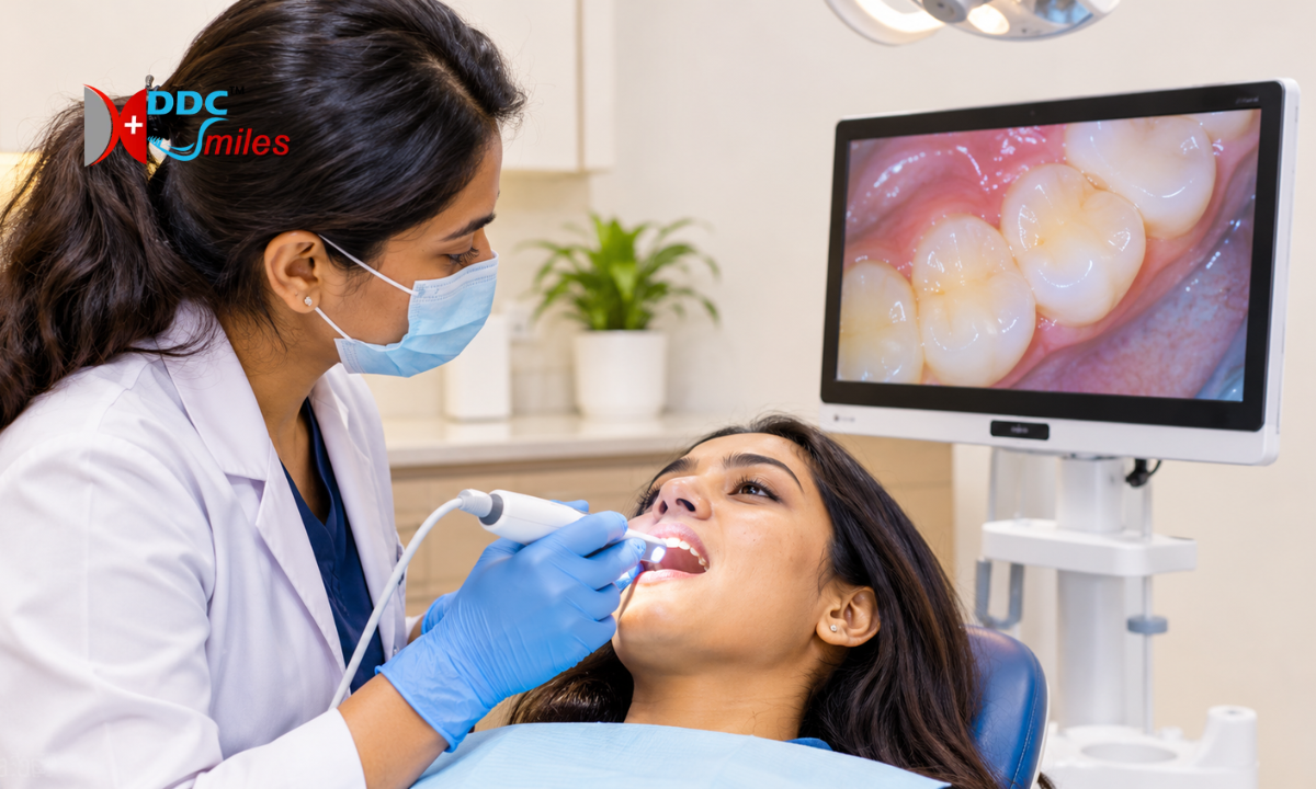
An intraoral camera in Bangalore helps dentists capture detailed images of the teeth, gums, and other visible areas inside the mouth. These images allow patients to see what the dentist observes during an examination and better understand recommended dental treatments.
At DDC Smiles, patients can enquire about dental examination and technology-assisted care at the clinics in Koramangala and HSR Layout, Bangalore. An intraoral camera supports visual examination, patient education, clinical documentation, and treatment planning. However, it does not replace a dentist’s examination or dental X-rays when additional diagnostic information is needed.
Looking for dental care in Bangalore? Contact DDC Smiles to enquire about an appointment at the Koramangala or HSR Layout clinic.
What Is an Intraoral Camera?
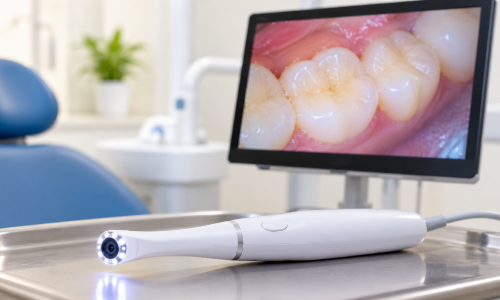
An intraoral camera is a small dental imaging device designed to capture photographs or video of the inside of the mouth. Its compact camera head allows the dentist to examine visible tooth surfaces, gum tissue, and areas that may be difficult to view directly.
The captured images can be displayed on a monitor, making it easier for patients to understand visible dental concerns and discuss possible treatment options.
Unlike a dental X-ray machine, an intraoral camera uses optical imaging rather than ionising radiation. It shows visible surfaces but cannot reveal all the structures beneath the teeth or gums.
How Does an Intraoral Dental Camera Work?
An intraoral dental camera captures images of selected areas inside the mouth. The dentist can view these images during the examination and explain any visible findings.
The process generally involves the following steps:
- Examination: The dentist examines your mouth and identifies areas that may benefit from closer visual assessment.
- Image capture: The camera is positioned to photograph selected teeth, gum tissue, or other visible areas.
- Display: The images are displayed on a screen when the equipment supports this function.
- Discussion: The dentist explains the visible findings and answers your questions.
- Further assessment: If necessary, the dentist may recommend dental X-rays or other clinical tests before confirming a diagnosis or treatment plan.
The process is generally straightforward and helps patients participate more actively in discussions about their oral health.
Benefits of Intraoral Camera Technology

An intraoral camera can improve communication between the dentist and patient by making visible dental conditions easier to understand.
1. Detailed Visual Examination
The camera provides magnified or close-up views of selected areas of the mouth, depending on the device. It can help the dentist examine visible tooth surfaces, gum tissue, existing restorations, and other areas of interest.
2. Better Patient Education
Dental conditions can be difficult to understand through verbal explanations alone. Viewing images on a screen can help patients understand what the dentist is discussing and why further assessment or treatment may be recommended.
3. Clinical Documentation
Intraoral photographs can help document visible oral conditions at a particular point in time. When comparable images are available from different appointments, the dentist may use them to review changes.
4. Support for Treatment Planning
Images can support discussions about restorative treatments, gum care, and other dental procedures. However, treatment planning also depends on clinical findings, patient history, symptoms, and any additional diagnostic tests required.
5. Improved Communication
Patients can ask questions while viewing the images, helping them make more informed decisions about their dental care.
What an Intraoral Camera Cannot Do
Although an intraoral camera is useful for visual examination, it has important limitations.
An intraoral camera cannot see beneath the tooth surface, inside the jawbone, or through the teeth in the way a dental X-ray can. It also cannot replace a clinical examination, patient history, or additional diagnostic tests when these are needed.
A normal-looking image does not rule out every dental problem. For example, decay between teeth, certain cracks, bone loss, and problems beneath existing restorations may require X-rays or further clinical assessment.
The dentist determines whether additional tests are necessary based on your symptoms, examination findings, and individual dental needs.
Intraoral Camera vs Dental X-Ray
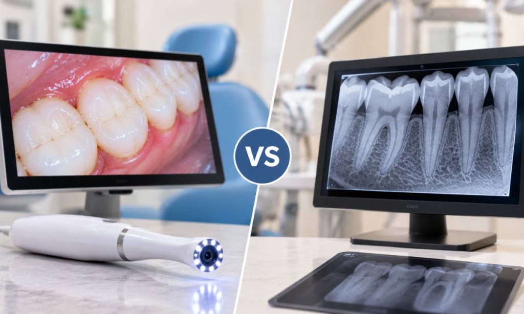
An intraoral camera and a dental X-ray provide different types of information. They can complement each other during a dental examination.
| Feature | Intraoral Camera | Dental X-Ray |
| Imaging method | Optical images of visible surfaces | Radiographic images using X-rays |
| Shows visible tooth surfaces | Yes | Not in the same photographic detail |
| Helps examine visible gum tissue | Yes | Limited |
| Shows tooth roots and internal structures | No | Yes, depending on the X-ray |
| Helps assess decay between teeth | May show visible changes, but cannot reliably assess all affected areas | Often useful |
| Shows bone structure | No | Yes, depending on the image |
| Uses ionising radiation | No | Yes |
| Main role | Visual examination, documentation, and patient education | Assessment of structures not fully visible during a clinical examination |
Your dentist will recommend the appropriate imaging method based on your dental condition. Not every patient needs an X-ray at every appointment.
When Might a Dentist Use an Intraoral Camera?
An intraoral camera may be used during a dental appointment when visual documentation or a closer view of a particular area would be helpful.
Examples include:
- Visible tooth damage: To document chips, worn surfaces, or other visible changes.
- Gum concerns: To capture images of visible gum tissue for discussion and documentation.
- Existing fillings and crowns: To examine accessible surfaces and document visible changes around restorations.
- Oral health education: To help explain plaque accumulation, visible staining, or other findings.
- Treatment discussions: To show patients the areas being considered for further examination or treatment.
- Monitoring visible changes: To compare images taken at different appointments when appropriate.
An intraoral camera does not confirm every dental condition on its own. The dentist interprets the images alongside the clinical examination and other relevant findings.
Is an Intraoral Camera Safe?
An intraoral camera uses light and optical imaging to capture pictures of the mouth. It does not expose patients to X-ray radiation.
The procedure is generally non-invasive and does not involve cutting or drilling. The camera is positioned to capture images of the areas being examined.
Appropriate infection-control procedures are also important. The intraoral camera should be used with suitable infection-control measures, including a protective barrier for the portion placed in the mouth and cleaning or disinfection of reusable surfaces between patients, following the manufacturer’s instructions and the clinic’s protocols.
If you have a sensitive gag reflex or feel uncomfortable during the examination, tell your dentist so the procedure can be adjusted where possible.
Dental Technology at DDC Smiles, Bangalore
Choosing a dental clinic involves more than the equipment used during an examination. A clear explanation of findings, an appropriate diagnostic process, and a treatment plan suited to your needs are also important.
At DDC Smiles, patients can enquire about dental services and the technology used to support examination and treatment planning.
Explore the dental services offered by DDC Smiles or learn more about the clinic’s dental facilities and technology.
Dental Clinic in Koramangala, Bangalore
Patients looking for a dental clinic in Koramangala, Bangalore, can contact DDC Smiles to enquire about appointments and available dental services.
An intraoral camera may help the dentist show visible dental findings and explain whether additional assessment is appropriate.
Visit the DDC Smiles website to learn more or use the contact page to enquire about an appointment.
Dental Clinic in HSR Layout, Bangalore
If you are looking for a dental clinic in HSR Layout, Bangalore, contact DDC Smiles to confirm appointment availability and the services offered at the clinic.
During your consultation, the dentist can assess your oral health, explain any visible findings, and recommend further tests when necessary.
For appointment enquiries, visit the DDC Smiles contact page.
Frequently Asked Questions About Intraoral Cameras
1. What is an intraoral camera used for?
An intraoral camera captures detailed images of visible areas inside the mouth. Dentists may use these images for visual examination, patient education, clinical documentation, and treatment discussions.
2. Does an intraoral camera detect cavities?
An intraoral camera may show visible signs of tooth decay, but it cannot detect every cavity. Decay between teeth or beneath the surface may require a clinical examination and dental X-rays.
3. Is an intraoral camera better than a dental X-ray?
Neither replaces the other because they provide different information. An intraoral camera shows visible tooth and gum surfaces, while X-rays help assess structures that cannot be seen directly. Your dentist may recommend one or both, depending on your needs.
4. Can an intraoral camera detect cracks in teeth?
It may help document cracks that are visible on the tooth surface. However, some cracks are difficult to identify with a camera alone. Depending on the examination, the dentist may use bite testing, transillumination, X-rays, or other clinical assessment methods.
5. Does an intraoral camera use radiation?
No. An intraoral camera uses optical imaging and does not produce ionising radiation. Dental X-rays use a different imaging method and are recommended when clinically appropriate.
6. Is an intraoral camera examination painful?
The procedure is generally non-invasive and does not usually cause pain. Some patients may experience mild discomfort if they have a sensitive gag reflex or difficulty keeping their mouth open.
7. Can intraoral photographs replace a dental examination?
No. Photographs provide useful visual information, but they cannot replace a comprehensive dental examination. The dentist may also need to review your symptoms, medical and dental history, and additional test results.
8. Where can I enquire about an intraoral camera examination in Bangalore?
You can contact DDC Smiles to enquire about dental appointments and the services available at its Koramangala and HSR Layout clinics. Visit the DDC Smiles contact page for appointment enquiries.
Book a Dental Consultation at DDC Smiles
An intraoral camera can help make dental examinations more informative by allowing patients to see visible findings and discuss them with their dentist. However, the right diagnosis and treatment plan depend on a complete clinical assessment and any additional tests required.
For dental appointment enquiries in Bangalore, contact DDC Smiles and ask about availability at the Koramangala or HSR Layout clinic.
References:
1)https://www.exportersindia.com/bangalore/dental-equipment.htm
2)https://pinkblue.in/agkem-intraoral-camera-with-wifi-1.html?srsltid=AYCX9vIYzV942Jxej-c4_wulDhb-5H10iuJzPO9gBmqQxs1thPmYfpi5
3)https://www.medicam.co.in/dental-camera.html
