

Tooth pain is often deceptive.
What feels like a simple toothache on the surface might actually be a severe infection brewing deep within the jawbone.
This hidden threat is clinically known as a Dentoalveolar Abscess.
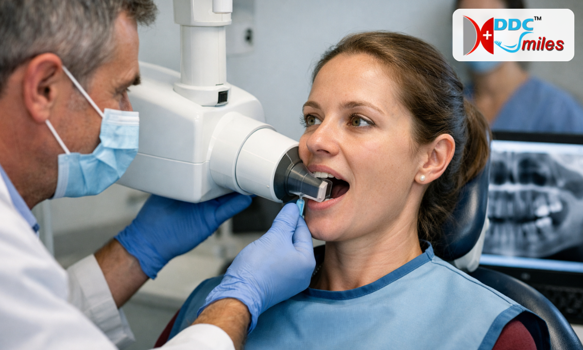
Patients often visit our clinics in Bangalore asking if we can simply drain the swelling without taking an X-ray.
The answer is almost always no.
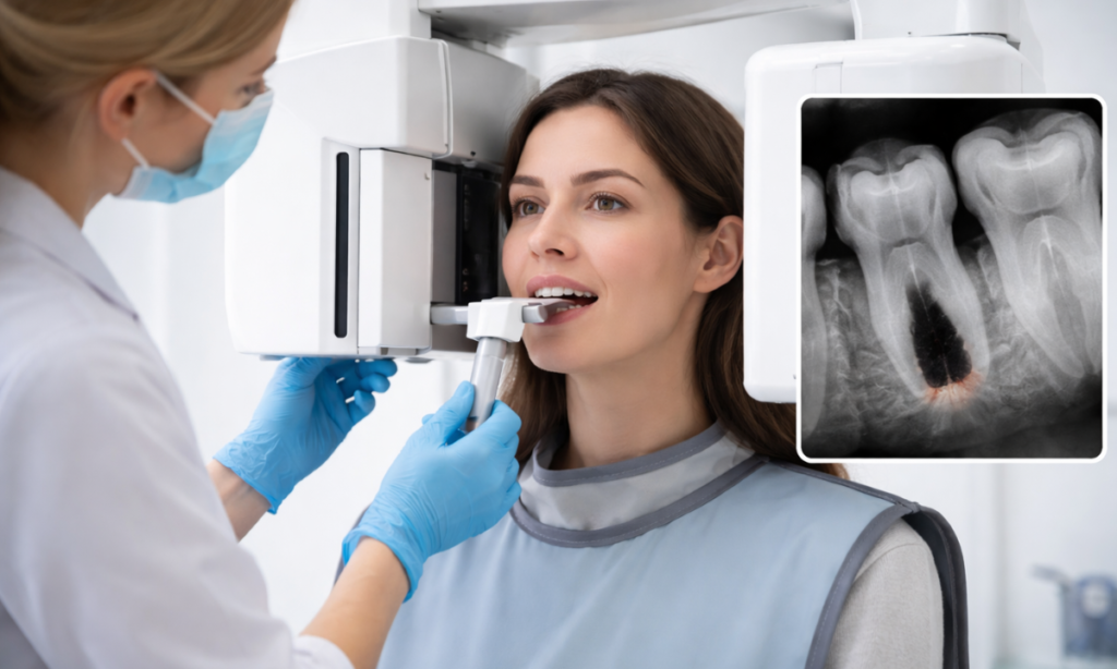
To treat the infection effectively, we must first see what the naked eye cannot. This is where a Dental Abscess Radiograph becomes the most critical tool in our diagnostic arsenal.
Understanding the Abscess Dental Definition is the first step toward relief.
In simple terms, a dental abscess is a pocket of pus caused by a bacterial infection. It can occur at different regions of the tooth, but the most common and painful type involves the root tip.
The Dental Abscesses Meaning goes beyond just pain.
It signifies that the bacteria have breached the tooth’s protective enamel, traveled through the root canal, and are now attacking the bone structure itself.
At DDC Smiles, we do not rely on guesswork.
Using advanced in-house diagnostic technology like RVG (Radio Visio Graphy) and CBCT (3D Scans), we capture precise images of the Periapical Abscess Radiograph.
This allows us to determine the exact extent of the infection and decide whether the tooth can be saved with a Root Canal Treatment or if it requires extraction to protect your overall health.
Medical Disclaimer
The information provided in this blog regarding Dental Abscess Radiograph protocols and treatments is for general educational purposes only. It does not constitute professional medical advice, diagnosis, or treatment.
Dentoalveolar Abscess and other oral infections can spread rapidly and require immediate clinical intervention. Do not use this article to self-diagnose or delay seeking professional care.
Always consult qualified dentists at DDC Smiles (Located in Koramangala and HSR Layout) for an accurate diagnosis.
Radiographic interpretations mentioned here are based on standard dental practices but may vary depending on the individual patient case.
What Is A Dentoalveolar Abscess? Understanding The Infection
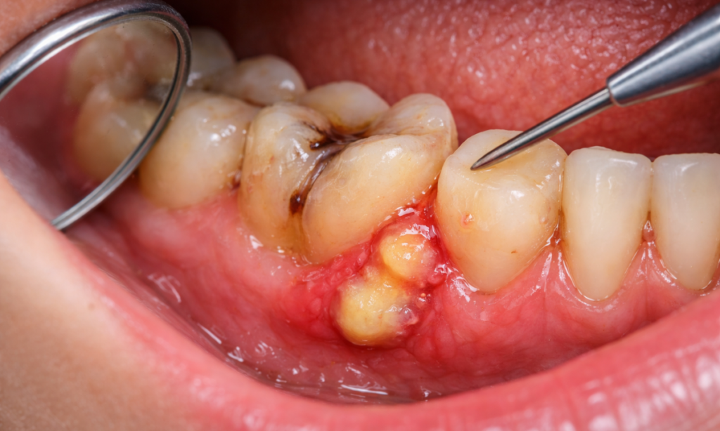
A Dentoalveolar Abscess is an accumulation of pus that forms inside the teeth, gums, or the bone that holds the teeth in place.
It is the body’s defensive reaction to a bacterial invasion.
The process usually begins with a simple cavity or a crack in the tooth.
If left untreated, bacteria penetrate the hard enamel and infect the soft pulp tissue inside.
This is where the nerves and blood vessels live. Once the pulp dies, the infection travels down the root canal and exits at the tip of the root, spreading into the surrounding bone.
This advanced stage is what we clinically term a Dentoalveolar Infection.
Types Of Dental Abscesses
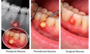
1. Periapical Abscess (Alveolar Abscess): This occurs at the tip of the tooth root. It is usually the result of an untreated cavity or a prior dental injury. The infection eats away at the bone, creating a hollow space filled with pus. This is the most common type requiring a Periapical Abscess Radiograph for diagnosis.
2. Periodontal Abscess (Gum Abscess): This occurs in the gums at the side of a tooth root. It is often caused by severe gum disease or a foreign object, like a popcorn hull getting stuck in the gum pocket.
3. Gingival Abscess: This is limited only to the gum tissue and does not affect the tooth or the periodontal ligament.
How To Identify Periapical Abscess: The Warning Signs
Patients often mistake a deep bone infection for a simple Cavity Abscess or sensitivity.
However, a true abscess presents specific symptoms that require immediate attention at DDC Smiles.
Checklist: Symptoms of a Developing Abscess
| Symptom | Description |
| Throbbing Pain | A continuous, intense pain that may radiate to the jawbone, neck, or ear. Lying down often makes it worse due to increased blood pressure in the head. |
| Temperature Sensitivity | Extreme pain when consuming hot or cold foods. The pain lingers even after the stimulus is removed. |
| Pain on Biting | Sharp pain when chewing or even tapping the tooth. This indicates the infection has spread from the tooth into the underlying bone or the alveolar abscess region. |
| Swelling | You may notice a pimple-like bump on your gum. If this ruptures, you might taste a foul-tasting fluid (pus) and feel immediate pain relief, but the infection remains active. |
| Systemic Signs | Fever, swollen lymph nodes under your jaw, or a general feeling of being unwell. |
If you experience these signs, the infection is no longer confined to the tooth. It has likely spread to the jawbone, making radiographic imaging mandatory to assess the damage.
Why Is Radiography Mandatory?
“Doctor, I can point to the tooth that hurts. Why do we need an X-ray?”
This is a common question at our front desk.
The reason is simple: We cannot treat what we cannot see. A visual examination only allows the dentist to see the Clinical Crown, the part of the tooth above the gum line.
However, a Periapical Abscess lives at the very bottom of the root, buried deep inside your jawbone.
The Iceberg Principle
Think of your tooth like an iceberg.
1. The Visible Tip: This is the white enamel you see when you smile.
2. The Submerged Mass: This is the root system and the surrounding bone, which makes up about 60% of the tooth structure.
In a Dentoalveolar Infection, the “explosion” happens at the bottom of the iceberg (the root tip).
By the time you see a swelling on the gum, the infection has likely already eaten through a significant amount of bone to reach the surface.
Without a Dental Abscess Radiograph, we are flying blind.
We would not know if the infection is small (treatable) or massive (requiring surgery).
What The X-Ray Reveals
When we take a radiograph at DDC Smiles, we are looking for specific Red Flags that are invisible to the naked eye:
1. Radiolucency: A dark shadow at the tip of the root. Healthy bone looks white; infected bone looks dark because the bacteria have destroyed the calcium.
2. Bone Loss: How much jawbone has been eaten away?
3. Proximity: Is the abscess dangerously close to your Sinus (upper jaw) or the Inferior Alveolar Nerve (lower jaw)?
4. Cysts: Has the abscess hardened into a radicular cyst?
A Note on Safety
Many patients hesitate due to fear of radiation. It is important to know that DDC Smiles uses Digital RVG (Radio Visio Graphy) technology, not old-school film.
1. Old Film X-Rays: Required higher radiation and chemical development.
2. Digital RVG: Uses a high-sensitivity sensor that requires 80% less radiation.
Perspective: The radiation exposure from a single dental X-ray is roughly equivalent to eating two bananas or taking a 2-hour flight. It is negligible and completely safe, even for children.
Types of Radiographs Used at DDC Smiles
At DDC Smiles (HSR Layout & Koramangala), we employ a tiered approach to imaging.
We do not just take random pictures; we select the specific type of Dental Abscess Radiograph that will give us the most information with the least radiation exposure.
Here are the three primary diagnostic tools we use to identify a Dentoalveolar Infection.
A. IOPA (Intra-Oral Periapical Radiograph)
This is the small sensor placed inside your mouth. It captures the entire tooth from the crown to the root tip and the surrounding bone.
1. Purpose: To detect a specific Periapical Abscess Radiograph pattern at the root apex of a single tooth.
2. Technology: We use RVG (Radio Visio Graphy) sensors. These digital sensors provide instant high-resolution images on our computer screens with minimal radiation.
3. Best For Confirming cavities, root infections, and checking bone levels between teeth.
B. OPG (Orthopantomogram)
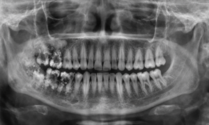
Sometimes, the infection is not limited to one tooth.
An OPG is a wide-angle X-ray that scans your entire upper and lower jaw, including all teeth, sinuses, and jaw joints, in a single image.
1. Purpose: To see if the Alveolar Abscess has spread to neighboring teeth or into the maxillary sinus (for upper teeth) or the mandibular nerve canal (for lower teeth).
Unlike many clinics that refer you to a diagnostic centre, we have an In-House OPG Machine. You get scanned and diagnosed immediately in the same appointment.
C. CBCT (Cone Beam Computed Tomography)
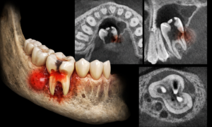
This is the gold standard for complex cases. A standard X-ray is 2-dimensional (flat), but your tooth is 3-dimensional.
1. Purpose: A CBCT scan allows us to slice the image layer by layer. We can see exactly how big the Cavity Abscess is, which direction it is spreading, and if there are hidden “accessory” canals in the root that a normal X-ray missed.
2. When It Is Required:
a. Failed Root Canals (Re-treatment).
b. Multi-rooted teeth (Molars) where roots overlap.
c. Planning for Dental Implants after extracting an infected tooth.
Table: Which Scan Do You Need?
| Scan Type | Coverage | Detail Level | Best For… |
| IOPA (RVG) | 1-2 Teeth | High (2D) | Routine Teeth Abscesses, Cavities, RCT Checks. |
| OPG | Full Jaw | Moderate (2D) | Multiple infections, Sinus issues, and wisdom teeth. |
| CBCT | Specific Area | Ultra-High (3D) | Complex Dentoalveolar Abscess, Failed RCTs, Implants. |
Interpreting the Image: What Are We Looking For?
When our specialists at DDC Smiles examine your Dental Abscess Radiograph, they are looking for specific changes in the bone density and root structure.
To the untrained eye, an X-ray might look like a blur of grey and white.
To a dentist, it tells the story of the infection’s timeline and severity.
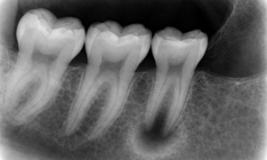
The Anatomy of a Shadow (Radiolucency)
The most obvious sign of a Dentoalveolar Infection is a change in color at the tip of the tooth root.
1. Healthy Bone: Appears light grey or white because it is dense and mineralized.
2. Infected Bone: Appears as a dark spot or “halo” around the root tip. We call this Radiolucency.
This dark area exists because the bacterial toxins from the Teeth Abscesses have dissolved the calcium in the jawbone. The larger the dark spot, the more bone has been destroyed.
The Early Warning Signs
Before a massive hole appears in the bone, there are subtle signs that a Periapical Abscess Radiograph will reveal.
1. Widening of the PDL Space: The Periodontal Ligament (PDL) is a thin black line that surrounds the root. In early infection, this line becomes thicker as the ligament swells due to inflammation.
2. Loss of Lamina Dura: This is the thin white line of dense bone that lines the tooth socket. If this white line disappears or becomes fuzzy at the root tip, it confirms that the infection has breached the socket wall.
Abscess vs. Cyst vs. Granuloma
Not all dark spots are simple abscesses. The shape of the radiolucency helps us classify the lesion.
1. Dental Abscess: Usually has irregular borders. It indicates an active, spreading infection.
2. Periapical Granuloma: A chronic reaction where the body tries to wall off the infection with granulation tissue. It appears as a well-defined round shadow.
If a granuloma is left untreated for years, it can liquefy and form a cyst. On a Dental Abscess Radiograph, this looks like a large, perfectly round dark circle with a distinct white outline.
Cysts often require surgical removal (Apicoectomy) in addition to Root Canal Treatment.
Treatment Options Based On The X-Ray Findings
Once the Periapical Abscess Radiograph confirms the location and severity of the infection, the treatment plan becomes clear.
At DDC Smiles, our priority is always to save the natural tooth whenever possible.
However, the X-ray is the final judge.
Here is how the radiographic evidence dictates your treatment options.
Root Canal Treatment (RCT)
If the X-ray shows that the Alveolar Abscess is localized around the root tip and the surrounding bone support is still strong, we proceed with a Root Canal Treatment.
1. The Procedure: We access the inside of the tooth, clean out the infected pulp and bacteria from the canals, and seal it.
2. The Goal: To remove the source of the Dentoalveolar Infection while keeping the natural tooth structure in your mouth.
We utilize Micro-Endodontics (Dental Microscopes). This allows us to see tiny accessory canals that standard vision misses, ensuring 100% of the infection is removed.
Tooth Extraction: When The Damage Is Severe
Sometimes, the Dental Abscess Radiograph reveals a grim picture.
If the infection has eaten away too much bone (Severe Bone Loss) or if the root has a vertical fracture extending below the gum line, the tooth cannot be saved.
1. The Procedure: The infected tooth is removed to stop the bacteria from spreading to the jawbone or bloodstream.
We can often place a Dental Implant immediately after extraction (Immediate Implants) to restore function and aesthetics without waiting months for healing.
Incision and Drainage (I&D)
If you come in with a massive, painful swelling on your face or gum, the X-ray might show a large accumulation of pus pressing against the tissue.
1. The Procedure: We make a small incision in the gum to drain the pus instantly. This provides immediate pain relief.
Important Note: This is not a cure. It is a temporary measure to relieve pressure. You will still need an RCT or Extraction afterward to fix the root cause.
A Critical Note on Antibiotics
Many patients ask: “Can I just take medicine to make it go away?”
The Answer is No.
Antibiotics travel through the blood. Since the inside of a dead tooth has no blood supply, the medicine cannot reach the bacteria hiding there.
Antibiotics can stop the swelling from spreading to your neck or eye, but they cannot cure a Teeth Abscesses.
Only physical treatment (RCT or Extraction) can eliminate the source.
Why Choose DDC Smiles For Abscess Treatment In HSR & Koramangala?
Precision Diagnostics Meets Expert Care.
When you are in the throes of a Dentoalveolar Infection, time is of the essence.
You do not want to visit a clinic, get a referral for a scan, travel to a diagnostic centre, and then come back the next day. Instead, you need a professional, fully equipped dental hospital in HSR Layout.
At DDC Smiles, we have built our practice around the philosophy of “Diagnosis First, Treatment Second.”
Here is why patients across Bangalore trust us with their complex dental emergencies:
1. The One-Stop Diagnostic Hub
Most dental clinics in Bangalore have to send patients out for advanced scans. DDC Smiles is one of the dental hospitals in Koramangala and HSR Layout, equipped with In-House OPG and CBCT (3D) Machines.
The Benefit: You walk in with pain, get your Dental Abscess Radiograph immediately, receive a diagnosis, and start treatment all in a single visit. No waiting, no traveling.
2. Hospital-Grade Sterilization
An abscess is an active bacterial infection. The last thing you need is a secondary infection from unsterile instruments. We operate as a Dental Hospital, not just a clinic.
1. Class B Autoclaves: We use the highest standard of sterilization for all surgical instruments.
2. Disposable Barriers: Every surface you touch is protected to prevent cross-contamination.
3. Micro-Endodontics
Dr. Jaikrishna and our team of Endodontists (Root Canal Specialists) use Dental Operating Microscopes.
1. The Difference: A standard eye sees the root canal as a tiny dot. A microscope sees it as a large tunnel.
We can find and clean hidden “accessory canals” that often cause Periapical Abscesses to return. This significantly raises the success rate of saving your natural tooth.
Conclusion
A Dentoalveolar Abscess is not a condition that goes away on its own. It is a ticking clock. The pain you feel is your body’s alarm system telling you that the infection is spreading.
1. Ignore it: You risk losing the tooth, damaging the jawbone, or spreading the infection to your face and neck.
2. Treat it: With a simple Dental Abscess Radiograph and timely intervention, we can eliminate the pain instantly and save your smile.
Do not guess with your health. If you have a throbbing toothache or a swelling on your gum, visit DDC Smiles today for a precise, digital diagnosis.
