
Securing an accurate maxillofacial diagnosis is the critical first step before any complex dental procedure.
Patients seeking a precise OPG X-ray in Bangalore require facilities equipped with advanced imaging infrastructure. Whether visiting a specialized dental clinic, superior diagnostic technology strictly dictates the success of the final treatment.
As a premier Dental Hospital in Koramangala and a leading Dental Hospital in HSR Layout, DDC Smiles utilizes state-of-the-art Digital OPG systems to provide unparalleled clinical clarity.
Standard intraoral bitewing X-rays only capture isolated, highly localized sections of the dental arch. However, comprehensive treatment planning for surgical implants, orthodontic realignment, or complex tooth extractions demands a complete, uninterrupted view of the entire maxillofacial region.
This strict anatomical requirement frequently leads patients to ask what an OPG X-ray is and how this specific scan alters their overall treatment trajectory.
Rather than merely outlining the procedure, understanding the profound clinical value of this diagnostic tool requires examining three specific dimensions of dental radiology:
1. The Technological Framework: Defining the exact extraoral imaging mechanics that allow a specialized machine to capture a continuous, flattened visualization of the complex, curved jawbone.
2. The Diagnostic Imperative: Outlining the specific maxillofacial pathologies, such as impacted third molars, hidden osseous cysts, and severe temporomandibular joint degradation, that strictly require panoramic evaluation before any surgical intervention.
3. The Procedural Protocol: Detailing the rapid, entirely noninvasive patient experience in the imaging suite and the subsequent pathological interpretation of the resulting two-dimensional data.
Thoroughly understanding the precise mechanical function and the diagnostic capabilities of a panoramic radiograph, patients can approach their advanced dental treatments with absolute clinical confidence.
What is an OPG X-ray?
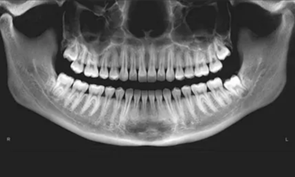
In clinical dentistry, an OPG, formally known as an Orthopantomogram, is a highly specialized extraoral radiological scan. Unlike standard intraoral X-rays, where the radiographic sensor is placed directly inside the patient’s mouth, the OPG utilizes advanced panoramic tomography.
This specific imaging modality allows the diagnostic team to capture the entire maxillofacial structure, encompassing the complete upper maxillary arch, the lower mandibular arch, all present dentition, and the complex temporomandibular joints, within a single continuous two-dimensional image.
To achieve this comprehensive visualization, the radiographic equipment relies on a highly synchronized mechanical process. The machine consists of two primary components mounted on a rotating arm: the X-ray emitter and the digital sensor.
During the scan, the emitter and the sensor rotate simultaneously in precisely opposite directions around the patient’s stationary head.
The clinical success of an OPG relies entirely on a critical radiological concept known as the focal trough. The focal trough is a specifically engineered, three-dimensional, horseshoe-shaped image layer programmed directly into the panoramic machine.
1. Within the Focal Trough: Any anatomical structure positioned precisely within this predefined three-dimensional zone, such as the primary dental arches and the supporting jawbones, appears sharply defined and clearly focused on the final digital radiograph.
2. Outside the Focal Trough: Any anatomical structure positioned physically outside of this specific diagnostic zone is intentionally blurred and rendered entirely out of focus by the rotating mechanics.
This highly specific tomographic blurring process eliminates visual interference from the dense bones of the cranial vault and the cervical spine.
It allows the dental surgeon to evaluate an unobstructed, flattened projection of the curved jawbone, providing the exact structural data strictly required for complex diagnostic evaluations.
A panoramic dental scan that provides a single, complete image of the entire oral region, including the upper and lower teeth, jawbones, wisdom teeth, temporomandibular joints, and surrounding sinus areas, offering a broader view than standard dental X-rays that capture only a few teeth at a time.
Conditions Identified via OPG Dental X-Ray
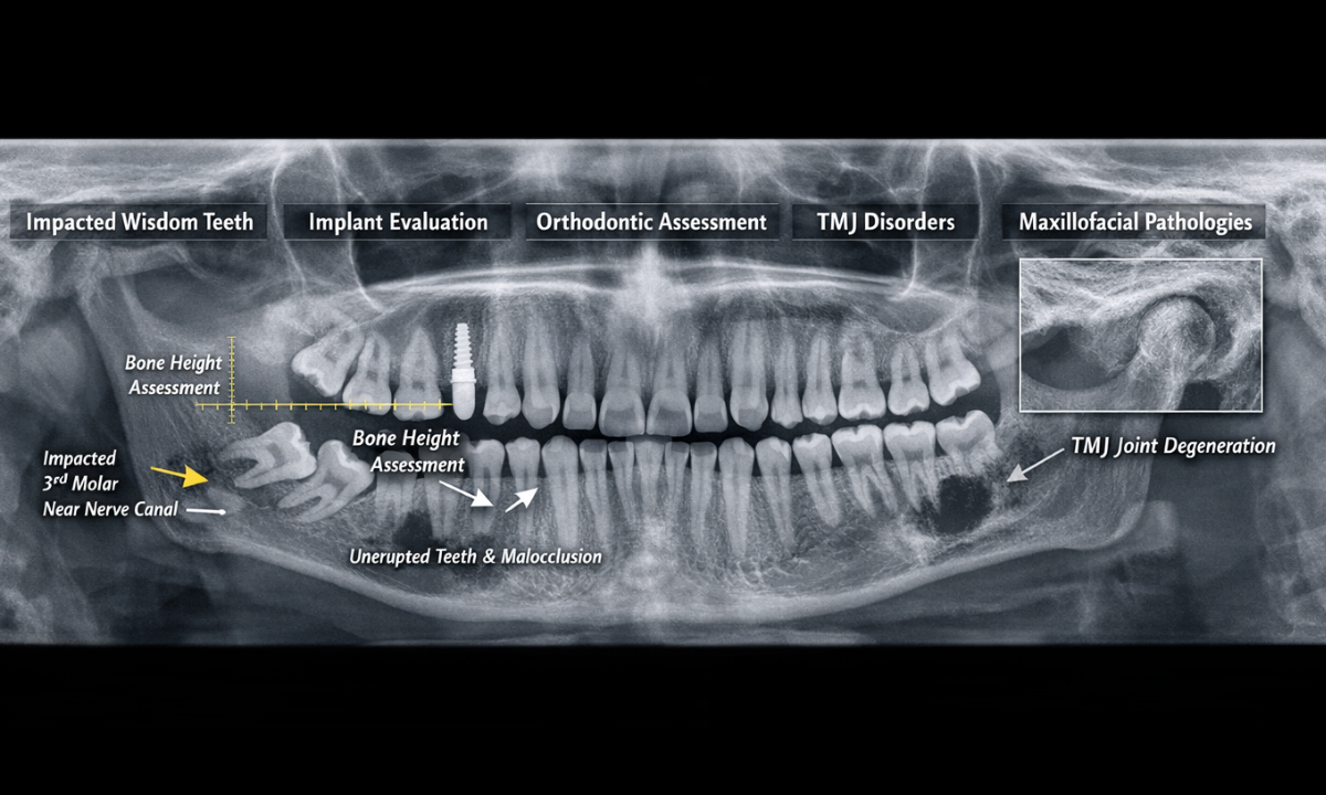
While standard intraoral radiographs are highly effective for detecting localized dental caries (cavities) or isolated pulpal infections, they are structurally incapable of providing a comprehensive maxillofacial overview. Maxillofacial surgeons, orthodontists, and specialized prosthodontists strictly require an OPG dental x-ray to evaluate the broader anatomical relationships within the jaw.
This scan is essential before performing complex dental procedures. By visualizing the entire lower facial skeleton simultaneously, the clinical team can definitively diagnose severe structural anomalies and carefully plan surgical interventions to minimize intraoperative risks.
The multidisciplinary dental team relies on panoramic imaging to identify and manage the following critical clinical conditions:
Primary Clinical Indications for Panoramic Radiography
| Clinical Pathology or Requirement | Targeted Anatomical Structure | Primary Diagnostic Objective of the OPG |
| Impacted Third Molars (Wisdom Teeth) | Mandibular angle and the inferior alveolar nerve canal. | To precisely determine the angle of tooth impaction and accurately measure the exact distance between the tooth roots and the primary mandibular nerve before complex surgical extraction. |
| Comprehensive Implant Planning | Maxillary and mandibular alveolar bone ridges. | To evaluate vertical bone height, assess overall bone density, and map the precise location of the maxillary sinuses to strictly ensure anatomical viability for titanium implant placement. |
| Advanced Orthodontic Assessment | The complete dental arch and all unerupted developing dentition. | To evaluate severe malocclusion, track the specific eruption pathways of permanent teeth in pediatric patients, and formulate precise, long-term structural alignment strategies. |
| Temporomandibular Joint (TMJ) Disorders | The temporomandibular joint condyles and the articular fossa. | To definitively detect gross structural asymmetry, advanced condylar degradation, or severe arthritic changes contributing to chronic jaw dysfunction and facial pain. |
| Hidden Maxillofacial Pathologies | Deep osseous tissues of the upper and lower jawbones. | To safely identify asymptomatic, deep-seated bone cysts, aggressive odontogenic tumors, or severe periodontal bone resorption that standard localized X-rays cannot capture. |
Acquiring this comprehensive radiological data, the dental surgeon effectively eliminates anatomical guesswork.
The resulting panoramic image provides the exact structural blueprint required to guarantee that all subsequent surgical or orthodontic treatments are executed with absolute clinical precision and maximum patient safety.
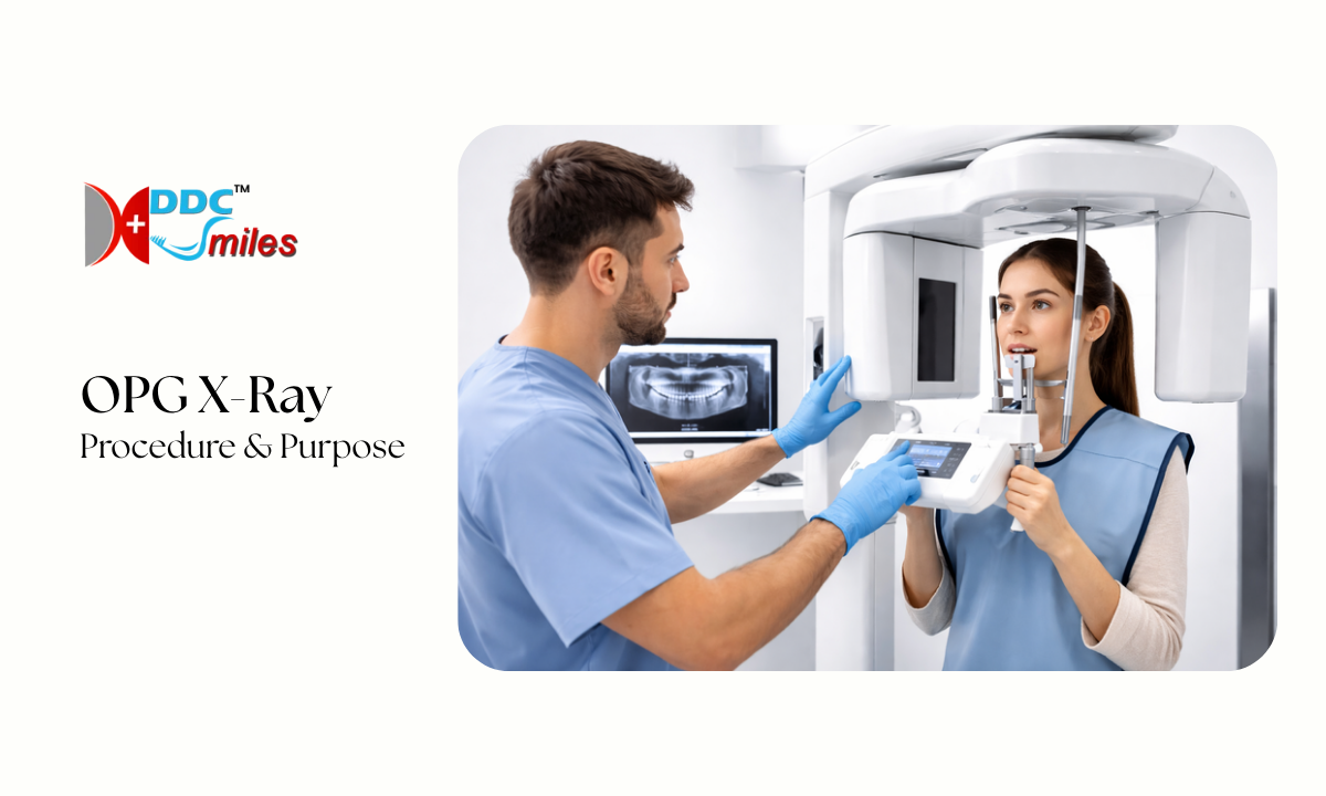
How is an OPG X-ray done?
Patients frequently experience apprehension before unfamiliar diagnostic procedures, often anticipating the physical discomfort associated with traditional intraoral sensors.
However, understanding exactly how an OPG X-ray is done immediately alleviates these clinical concerns. The panoramic imaging procedure is entirely extraoral, completely noninvasive, painless, and exceptionally fast.
The entire diagnostic process is conducted on an outpatient basis and follows a highly structured, strict clinical sequence:
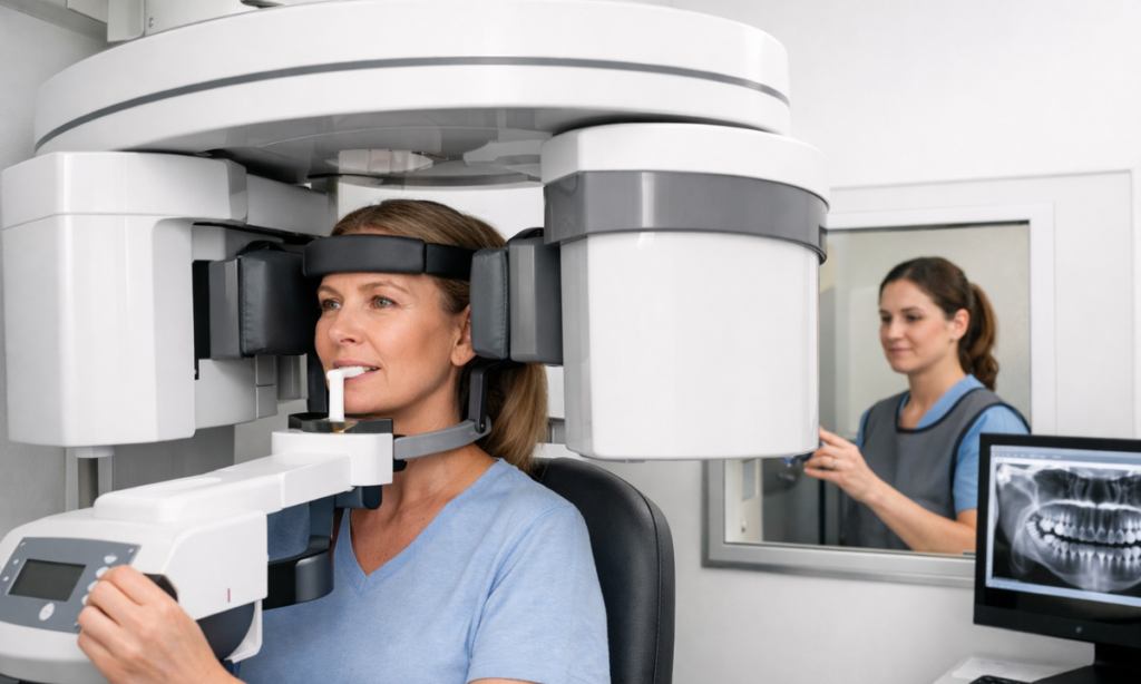
1. Pre-Procedural Preparation: The radiologic technologist will instruct the patient to physically remove all metallic objects from the head and neck region.
This strictly includes earrings, necklaces, facial piercings, eyeglasses, hearing aids, and any removable dental prosthetics. High-density metallic materials physically block the radiation beam, creating severe white radiological artifacts on the final image that completely obscure the underlying anatomical structures.
2. Anatomical Positioning: The patient is guided to stand or sit squarely within the center of the panoramic unit. The technologist precisely adjusts the vertical height of the machine to align with the patient.
The patient then rests their chin on a specialized physical support and gently bites down on a small, sterile plastic bite peg. This exact positioning is strictly mandatory. It physically separates the upper and lower teeth and aligns the dental arches perfectly within the machine’s predefined focal trough.
3. Cranial Stabilization: To prevent any inadvertent micro movements that would severely degrade the final image resolution.
The technologist gently secures the patient’s head utilizing padded lateral temple supports and an anterior forehead rest.
4. The Active Imaging Phase: The technologist steps behind a lead shielded barrier to manually initiate the scan. The mechanical arm of the OPG unit continuously rotates in a synchronized semicircle around the patient’s head.
This active scanning phase requires the patient to remain completely motionless and hold their breath for approximately ten to fifteen seconds.
5. Digital Acquisition: Once the mechanical rotation concludes, the machine instantly ceases all radiation output.
As modern facilities utilize digital sensors rather than chemical film, the high-resolution two-dimensional image is immediately transmitted to the clinic’s computer network for instant pathological review by the dental surgeon.
This simple, noninvasive process ensures accurate imaging while keeping patients comfortable and radiation exposure minimal.
Analyzing OPG X-Ray Images and Cost
When a maxillofacial radiologist or dental surgeon reviews an OPG X-ray image, they are evaluating a flattened, two-dimensional projection of a complex, curved three-dimensional structure.
The resulting digital radiograph presents the entire lower facial skeleton as a continuous, wide U shape. The clinician does not simply look at the teeth. Instead, they systematically analyze several distinct anatomical zones to formulate a comprehensive, highly accurate diagnosis.
Anatomical Landmarks Evaluated in a Panoramic Radiograph
| Targeted Anatomical Zone | Visual Representation on the OPG | Primary Clinical Significance |
| The Complete Dental Arches | Displays all erupted and completely unerupted dentition in a single, continuous panoramic row. | Identifies advanced interproximal decay, overall root angulation, congenitally missing teeth, and the precise impaction angles of wisdom teeth. |
| The Maxillary Sinuses | Appears as large, dark, radiolucent anatomical cavities positioned directly above the upper posterior teeth. | Strictly critical for determining the exact available vertical bone height before authorizing maxillary titanium implant placement. |
| The Mandibular Canal | Seen as a distinct dark channel running horizontally through the dense lower jawbone. | Houses the inferior alveolar nerve. Mapping its precise anatomical location prevents permanent neurological damage during complex surgical extractions. |
| The Temporomandibular Joints (TMJ) | Located at the extreme upper left and upper right peripheral edges of the digital scan. | Evaluates the bony articular condyles for severe arthritic degradation, gross structural asymmetry, or traumatic physical dislocation. |
Beyond the clinical interpretation of the data, patients actively planning their treatments frequently inquire about the financial aspect, specifically asking how much an OPG X-ray costs.
In Bangalore, the pricing for this highly advanced diagnostic scan remains relatively standardized and accessible. The cost for a high-resolution digital OPG typically ranges between ₹600 and ₹1,200 INR. This specific price variance depends entirely on the generation of the digital sensor utilized and the overall technological infrastructure of the diagnostic facility.
While isolated external scan centers offer this imaging service, undergoing the procedure directly within a fully equipped dental hospital provides a distinct clinical advantage. It eliminates the requirement for secondary travel appointments, allows the operating surgeon to review the digital data instantly, and accelerates the transition directly into active treatment planning.
What is Dental CBCT and how is it Different from an OPG?
While an OPG provides a comprehensive two-dimensional overview of the entire maxillofacial region, certain complex dental conditions demand an even higher level of diagnostic precision. This is where Dental CBCT (Cone Beam Computed Tomography) becomes clinically indispensable.
Dental CBCT is an advanced three-dimensional imaging modality that captures highly detailed volumetric data of the teeth, jawbone, nerves, and surrounding anatomical structures. Unlike the flattened projection seen in an OPG, CBCT generates a precise 3D reconstruction of the patient’s oral and maxillofacial anatomy.
This enhanced imaging capability allows clinicians to evaluate structures layer by layer with absolute spatial accuracy.
Key Clinical Advantages of Dental CBCT:
1. Three-Dimensional Visualization: Provides a complete 3D view of bone, teeth, and soft tissue relationships, eliminating the limitations of 2D imaging.
2. Accurate Implant Planning: Enables precise measurement of bone width, height, and density, ensuring optimal placement of dental implants.
3. Nerve Mapping: Clearly identifies the exact position of critical structures like the inferior alveolar nerve, significantly reducing surgical risks.
4. Complex Pathology Detection: Helps diagnose hidden infections, cysts, tumors, and fractures with exceptional clarity.
5. Endodontic Precision: Assists in detecting root canal complexities, fractures, and previously missed canals.
OPG vs CBCT – Clinical Perspective:
1. OPG: Ideal for initial screening, orthodontic planning, and evaluating overall jaw structure.
2. CBCT: Recommended for advanced surgical procedures, implantology, and cases requiring detailed anatomical mapping.
At DDC Smiles, both OPG and CBCT technologies are strategically integrated to ensure that every diagnosis is supported by the most appropriate imaging modality. While an OPG serves as the foundational diagnostic scan, CBCT is selectively utilized when absolute three-dimensional accuracy is required for complex dental interventions.
Why Choose DDC Smiles?
We fundamentally understand that exceptional dental treatment begins strictly with flawless clinical diagnostics. With a trusted medical legacy spanning over two decades, our specialized facilities eliminate the traditional, time-consuming inconvenience of outsourced radiological imaging.
By integrating advanced diagnostic technology directly into our clinical workflow, we provide our patients with several distinct medical advantages:
1. In-House Panoramic Infrastructure: Both our Koramangala and HSR Layout branches feature state-of-the-art Digital OPG and 3D Cone Beam Computed Tomography systems directly on site. This guarantees immediate image acquisition and prevents patients from navigating external diagnostic centers while in severe dental pain.
2. Expert Clinical Interpretation: Under the specialized medical leadership of Dr. H. J. Jaikrishna, our multidisciplinary team instantly analyzes your high-resolution panoramic data. This immediate review allows our surgeons to formulate highly precise, personalized treatment strategies during your very first consultation.
3. Uncompromising Safety Standards: We rigorously adhere to a strict 5-level sterilization protocol within our dedicated imaging suites. This ensures maximum patient safety and absolute cross-infection control during every single diagnostic procedure.
Choosing DDC Smiles means selecting a comprehensive dental hospital in Bangalore where advanced diagnostics and elite surgical execution operate seamlessly under one roof.
Conclusion
An OPG scan is not merely a supplementary X-ray; it is the definitive structural blueprint strictly required for advanced maxillofacial interventions. By providing a comprehensive, uninterrupted visualization of the entire lower facial skeleton, this rapid, noninvasive technology empowers dental surgeons to execute complex procedures with absolute anatomical safety and clinical predictability.
If you require an advanced orthodontic assessment, are considering titanium dental implants, or need a surgical evaluation for impacted wisdom teeth, contact DDC Smiles today. Schedule your comprehensive diagnostic consultation and experience the distinct clinical advantage of integrated, highly advanced dental care.
