
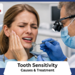
Patients often panic when they notice a sudden shift in their oral tissue tone.
Seeking immediate professional evaluation is critical. A specialized dental hospital in HSR Layout and Koramangala provides the exact diagnostics needed to identify the underlying cause.
Delaying an assessment allows potential periodontal infections to spread rapidly. Residents actively searching for reliable gum treatment in HSR layout require clinical clarity rather than internet speculation.
You must understand the biological mechanisms driving this physical change.
The discoloration of gums stems from a wide variety of physiological and pathological sources. Assessing the gums normal color is the mandatory first step.
Healthy gingival tissue does not conform to a single, universal shade of pink.
It varies significantly based on individual genetics, blood flow, and epithelial thickness.
When patients observe their gums color change from light pink to deep brown, purple, or black, the shift indicates a structural or chemical alteration at the cellular level. Determining exactly what causes gum discoloration dicta
Genetics and Melanin
The human body produces a specialized pigment called melanin.
This complex polymer determines the exact shade of your hair, skin, and eyes. It performs the same function inside your mouth.
Specialized cells known as melanocytes reside in the basal layer of the gingival epithelium. These cells continuously manufacture melanin granules and distribute them to the surrounding tissue.
Evaluating the normal color requires looking at a patient’s ethnic background. Individuals with darker skin tones naturally possess highly active melanocytes.
Their gingival tissue will reflect this genetic trait immediately.
Table: The Spectrum of Healthy Gingival Tissue
| Tissue Appearance | Melanin Activity Level | Clinical Status |
| Light Coral Pink | Very Low | Perfectly healthy baseline, typically seen in lighter skin tones. |
| Light Brown to Purple | Moderate | Perfectly healthy baseline, frequently observed in medium skin tones. |
| Deep Brown to Black | High | Perfectly healthy baseline, naturally occurring in darker skin tones. |
Dentists classify this natural darkening as physiologic pigmentation. It is a completely benign anatomical feature.
A gums color black appearance in a patient of African, Asian, or Mediterranean descent rarely indicates active disease. The dark coloration usually presents symmetrically across both the upper and lower dental arches.
To differentiate between a genetic trait and a sudden medical issue, clinicians look for specific physical markers.
1. Symmetry: Genetic pigmentation is distributed evenly across the entire mouth.
2. Tissue Texture: Naturally dark gums retain a firm, stippled surface resembling an orange peel.
3. Age of Onset: Physiological color establishes itself distinctly during the first two decades of life.
Suddenly, localized dark spots appearing rapidly in adulthood defy these natural patterns. Asymmetrical, flat black lesions require immediate clinical biopsy to definitively rule out malignant mucosal melanoma.
External Factors: What Causes Gum Discoloration
Identifying exactly what causes gum discoloration in adulthood requires looking past genetics.
External environmental factors and clinical interventions frequently alter tissue pigmentation. A sudden gum color change often points directly to lifestyle habits or previous dental work.
The most common environmental trigger is tobacco use.
Medical professionals classify this specific reaction as Smoker’s Melanosis. Tobacco smoke contains thousands of toxic chemicals.
Nicotine directly stimulates the melanocytes resting in the gum tissue. The body aggressively produces extra melanin as a biological defense mechanism against chronic heat and chemical toxins.
This results in a diffuse, patchy darkening, typically appearing prominently on the lower front gums. Quitting smoking often allows the tissue to gradually return to its normal shade over several years.
Previous dental procedures also leave permanent marks. Amalgam tattoos represent a very frequent, entirely harmless cause of localized darkening.
Old silver dental fillings contain a mixture of metals.
During the placement, polishing, or removal of these traditional restorations, microscopic metallic particles can accidentally embed themselves into the surrounding mucosal tissue.
The gums heal completely over these fragments. This trapped metal oxidizes over time, creating a distinct, flat, blue, gray, or gum-colored black patch directly adjacent to the treated tooth.
Systemic medications also induce notable pigmentation changes as a direct pharmacological side effect.
1. Minocycline: Dermatologists frequently prescribe this heavy-duty antibiotic for severe acne. It chemically binds to underlying bone and tooth roots, casting a permanent blue-gray shadow through the translucent gum tissue.
2. Antimalarial Drugs: Medications such as chloroquine directly stimulate pigment production across the entire oral mucosa.
3. Heavy Metal Exposure: Chronic occupational or environmental exposure to lead, bismuth, or mercury creates a highly distinct, dark line precisely at the gingival margin.
Table: External Triggers and Their Clinical Appearance
| External Factor | Typical Location | Visual Characteristics |
| Tobacco Smoke | Lower front gums, inner cheeks. | Diffuse, irregular brown or black patches. |
| Amalgam Particles | Next to a tooth with a silver filling. | Small, isolated, flat blue or gray spot. |
| Systemic Medications | Widespread across the upper and lower arches. | Generalized blue, gray, or brown tinting. |
Recognizing these external triggers immediately prevents unnecessary patient panic.
A dentist can easily diagnose an amalgam tattoo by taking a standard dental X-ray to spot the radiopaque metal fragments. This simple diagnostic step definitively separates a harmless metallic stain from a serious, aggressive pathological lesion.
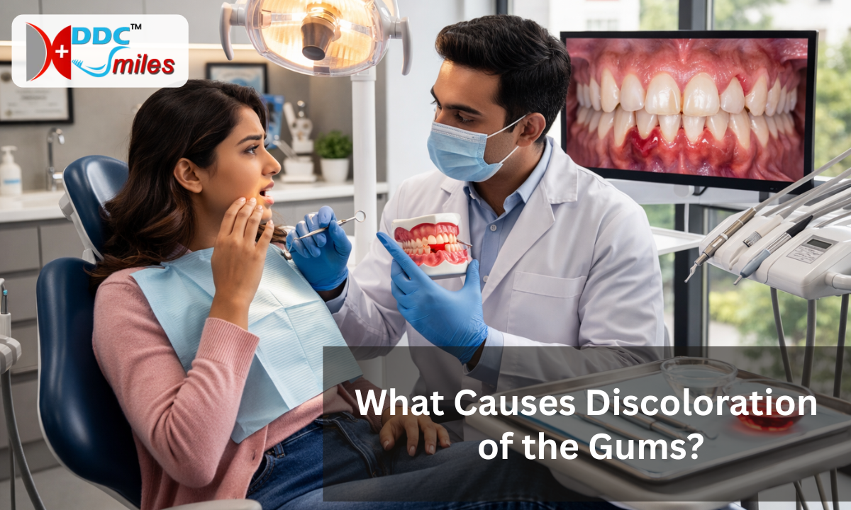
Clinical Conditions and Gum Disease
Pathological infections demand immediate attention.
When aggressive oral bacteria penetrate beneath the gum line, the gingival tissue physically reacts to the invasion. A sudden gums color change often signals severe underlying disease rather than a harmless surface stain.
Acute Necrotizing Ulcerative Gingivitis (ANUG) represents a severe, painful bacterial infection. Medical professionals historically refer to this condition as trench mouth.
The aggressive bacteria rapidly destroy the interdental papillae, which are the delicate triangular gum peaks between your teeth. As this specific tissue actively dies and necrotizes, it turns a distinct grayish-black.
Chronic periodontal disease presents a different visual signature.
The persistent bacterial plaque triggers massive localized inflammation. The human body floods the infected area with excess blood to fight the active bacterial colonies.
This chronic vascular engorgement transforms the tissue from a healthy pink into a swollen, deep purple or dark red shade.
Systemic medical conditions also manifest directly inside the oral cavity.
1. Addison’s Disease: This rare endocrine disorder causes severe adrenal gland failure.
The pituitary gland overcompensates by pumping out massive amounts of adrenocorticotropic hormone (ACTH).
This specific hormone directly overstimulates the melanocytes, creating striking, dark brown or black patches across the mucosal lining.
2. Peutz-Jeghers Syndrome: This genetic condition causes benign polyps in the gastrointestinal tract.
It simultaneously triggers the formation of distinct, dark blue or brown freckle-like spots on the lips and gums.
3. Oral Malignant Melanoma: This is an aggressive, dangerous form of mucosal cancer.
It frequently appears on the upper maxillary gums as an asymmetrical, rapidly expanding dark lesion with highly irregular borders.
Differentiating between a localized vascular inflammatory response and a systemic endocrine disorder requires specialized diagnostic training.
A dentist must physically probe the tissue, evaluate the depth of the periodontal pockets, and review your complete medical history to accurately diagnose the exact pathology driving the discoloration.
Treatment for Gum Discoloration
When patients seek a treatment for gum discoloration, the clinical approach depends entirely on the initial diagnosis.
Cosmetic masking fails if an active bacterial infection is destroying the supporting bone.
A dentist must cure the underlying pathology before addressing the superficial pigmentation.
If periodontitis or ANUG drives the tissue darkening, the protocol is strictly antimicrobial.
1. Deep Scaling and Root Planning: The hygienist physically scrapes the hardened bacterial calculus from deep beneath the gum line.
Removing this infectious reservoir halts the severe vascular inflammation. The swollen, dark purple tissue gradually shrinks and returns to a healthy, oxygenated pink state.
2. Targeted Antibiotic Therapy: Severe necrotic infections require systemic pills or localized antibacterial rinses to kill the aggressive anaerobic bacteria rapidly.
Cosmetic darkening from excess melanin requires a completely different surgical approach.
The pigment sits securely in the superficial layers of the epithelium. Dentists physically strip away this microscopic outer layer to expose the lighter connective tissue underneath.
3. Laser Depigmentation: This is the modern clinical standard for cosmetic lightening. The periodontist uses a precise dental diode laser to ablate the active melanocytes.
The targeted light energy vaporizes the pigment and cauterizes the microscopic blood vessels simultaneously. This advanced technique ensures minimal bleeding and remarkably fast healing.
4. Scalpel Gingivectomy: Traditional surgical peeling physically slices away the hyperpigmented outer tissue layer.
The human body naturally regenerates a new, unpigmented epithelial shield over several weeks.
5. Rotary Microabrasion: A specialized, high-speed dental bur physically sands away the pigmented epithelial cells.
Selecting the correct surgical modality requires evaluating the gingival thickness and the exact physical depth of the pigment.
Eradicating a deep amalgam tattoo often requires a minor soft tissue graft to replace the permanently stained mucosa completely. The clinical goal always prioritizes preserving the structural integrity of the tooth root over achieving an artificial cosmetic shade.
Why Choose DDC Smiles
Diagnosing a sudden gingival color shift requires specialized periodontic expertise.
Masking a severe bacterial infection with cosmetic lasers is clinical malpractice. At DDC Smiles, our specialists prioritize foundational oral health over superficial aesthetics.
We actively investigate the biological root cause of your specific tissue changes.
1. Pathology First: Our diagnostic protocol mandates strict screening for aggressive necrotizing bacteria and heavy metal toxicity before we approve any cosmetic depigmentation procedures.
2. Advanced Laser Therapy: We operate precision dental diode lasers to perform safe, highly targeted melanin ablation with minimal recovery time.
3. Comprehensive Biopsies: Our clinical team immediately isolates and tests asymmetrical, rapidly expanding dark lesions to definitively rule out malignant mucosal melanoma.
Your custom treatment plan relies entirely on an exact microscopic diagnosis.
This clinical rigor ensures we permanently resolve the underlying pathology rather than temporarily painting over a spreading infection.
Conclusion
Oral tissue acts as a highly sensitive mirror for systemic and localized disease.
A sudden shift from a normal pink baseline to a deep purple or black shade demands immediate clinical evaluation.
Genetics firmly dictate your natural melanin levels. Rapid, localized color changes usually signal active bacterial destruction, pharmacological side effects, or embedded foreign materials.
Do not wait for the gingival tissue to actively necrotize or recede.
Schedule a diagnostic periodontal evaluation at DDC Smiles today. Our clinical experts will physically map your exact gingival architecture and deploy the precise antimicrobial or surgical protocols required to restore your oral health entirely.
